

2.2.6 FIB-Milling
for Imaging Thicker
Regions of the Cell
In cryo-ET, sample thickness is a critical factor for data collection.
Because of inelastic scattering, the application of cryo-ET is usually
restricted to samples thinner than 600 nm, like small bacterial cells
or the thin peripheral regions of eukaryotic cells. However, most
regions of eukaryotic cells and many bacteria are thicker than that;
thus, additional thinning procedures are required for cryo-ET to
gain access to these samples (Fig. 2a). In recent years, cryo-FIBmilling has been developed as a tool to make thin lamellae from a
thick sample while preserving most of the native state of the molecules. The concept of FIB-milling is to use high-energy ions such as
gallium to remove materials above and below a targeted area. When
milling at a low angle, this will create a lamella, usually a few
hundred nanometers thick amenable for cryo-ET (Fig. 2b–d).
Here, we describe how to do cryo-FIB-milling on an FEI Versa
FIB/SEM equipped with a Quorum cryo-transfer system.
1. To prepare the Quorum transfer system for cryo work, mount
the Quorum cryo-stage on the SEM stage.
2. Start the Quorum system in manual mode to pump vacuum in
the chamber and scaffold. Prep-chamber vacuum should reach
less than 6 Â 10
À6 before cooling to avoid contamination.
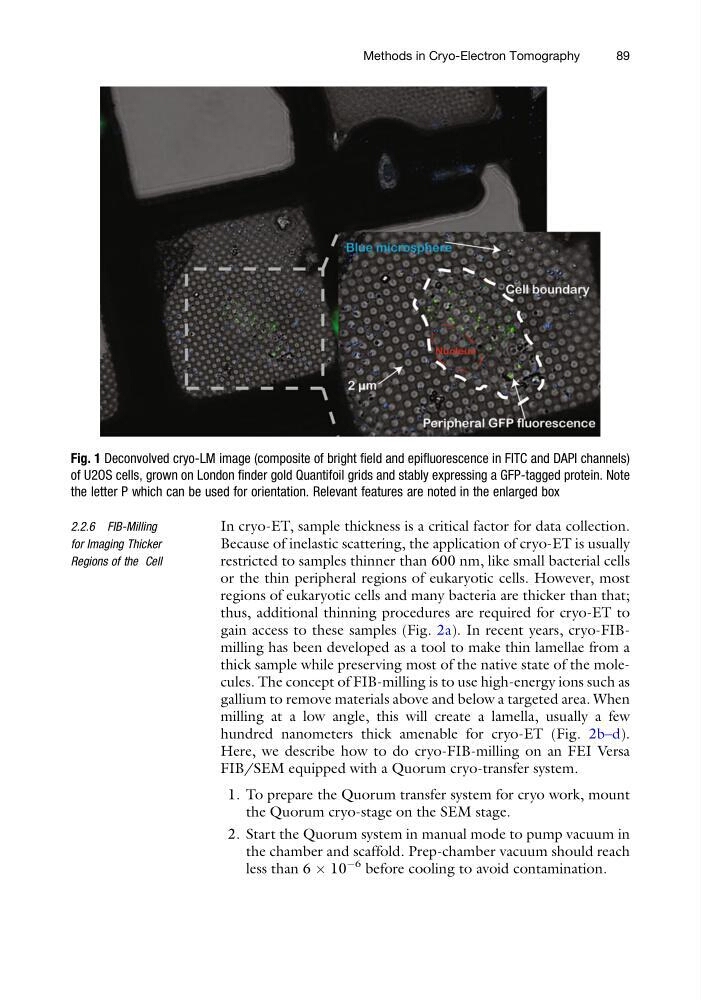
Fig. 1 Deconvolved cryo-LM image (composite of bright field and epifluorescence in FITC and DAPI channels)
of U2OS cells, grown on London finder gold Quantifoil grids and stably expressing a GFP-tagged protein. Note
the letter P which can be used for orientation. Relevant features are noted in the enlarged box
Methods in Cryo-Electron Tomography
89
for Imaging Thicker
Regions of the Cell
In cryo-ET, sample thickness is a critical factor for data collection.
Because of inelastic scattering, the application of cryo-ET is usually
restricted to samples thinner than 600 nm, like small bacterial cells
or the thin peripheral regions of eukaryotic cells. However, most
regions of eukaryotic cells and many bacteria are thicker than that;
thus, additional thinning procedures are required for cryo-ET to
gain access to these samples (Fig. 2a). In recent years, cryo-FIBmilling has been developed as a tool to make thin lamellae from a
thick sample while preserving most of the native state of the molecules. The concept of FIB-milling is to use high-energy ions such as
gallium to remove materials above and below a targeted area. When
milling at a low angle, this will create a lamella, usually a few
hundred nanometers thick amenable for cryo-ET (Fig. 2b–d).
Here, we describe how to do cryo-FIB-milling on an FEI Versa
FIB/SEM equipped with a Quorum cryo-transfer system.
1. To prepare the Quorum transfer system for cryo work, mount
the Quorum cryo-stage on the SEM stage.
2. Start the Quorum system in manual mode to pump vacuum in
the chamber and scaffold. Prep-chamber vacuum should reach
less than 6 Â 10
À6 before cooling to avoid contamination.
Fig. 1 Deconvolved cryo-LM image (composite of bright field and epifluorescence in FITC and DAPI channels)
of U2OS cells, grown on London finder gold Quantifoil grids and stably expressing a GFP-tagged protein. Note
the letter P which can be used for orientation. Relevant features are noted in the enlarged box
Methods in Cryo-Electron Tomography
89