

lysine to facilitate cell adhesion. If sterility is important, grids may
be sterilized under a germicidal UV lamp prior to coating and
seeding with cells. The appropriate seeding concentration and
recovery periods should be determined empirically.
Some mammalian cells may be deposited on grids immediately
before plunging, similarly to protein solutions for single-particle
cryo-EM. In this case, adherent cells may be dissociated from the
culture surface (using trypsin/EDTA) with the aim to create single
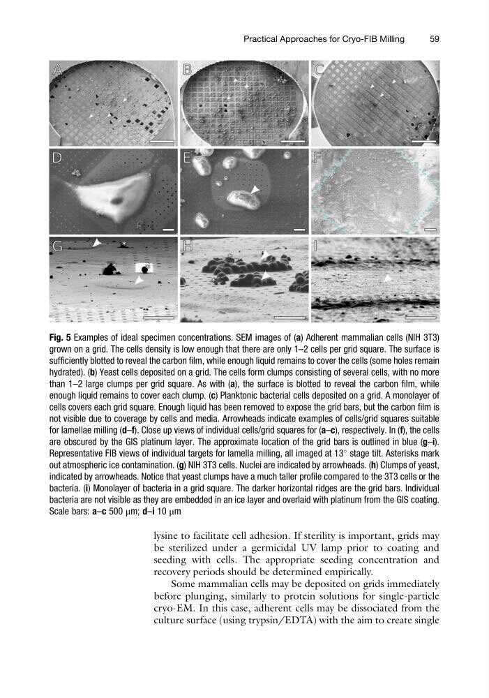
Fig. 5 Examples of ideal specimen concentrations. SEM images of (a) Adherent mammalian cells (NIH 3T3)
grown on a grid. The cells density is low enough that there are only 1–2 cells per grid square. The surface is
sufficiently blotted to reveal the carbon film, while enough liquid remains to cover the cells (some holes remain
hydrated). (b) Yeast cells deposited on a grid. The cells form clumps consisting of several cells, with no more
than 1–2 large clumps per grid square. As with (a), the surface is blotted to reveal the carbon film, while
enough liquid remains to cover each clump. (c) Planktonic bacterial cells deposited on a grid. A monolayer of
cells covers each grid square. Enough liquid has been removed to expose the grid bars, but the carbon film is
not visible due to coverage by cells and media. Arrowheads indicate examples of cells/grid squares suitable
for lamellae milling (d–f). Close up views of individual cells/grid squares for (a–c), respectively. In (f), the cells
are obscured by the GIS platinum layer. The approximate location of the grid bars is outlined in blue (g–i).
Representative FIB views of individual targets for lamella milling, all imaged at 13
stage tilt. Asterisks mark
out atmospheric ice contamination. (g) NIH 3T3 cells. Nuclei are indicated by arrowheads. (h) Clumps of yeast,
indicated by arrowheads. Notice that yeast clumps have a much taller profile compared to the 3T3 cells or the
bacteria. (i) Monolayer of bacteria in a grid square. The darker horizontal ridges are the grid bars. Individual
bacteria are not visible as they are embedded in an ice layer and overlaid with platinum from the GIS coating.
Scale bars: a–c 500 μm; d–i 10 μm
Practical Approaches for Cryo-FIB Milling
59
be sterilized under a germicidal UV lamp prior to coating and
seeding with cells. The appropriate seeding concentration and
recovery periods should be determined empirically.
Some mammalian cells may be deposited on grids immediately
before plunging, similarly to protein solutions for single-particle
cryo-EM. In this case, adherent cells may be dissociated from the
culture surface (using trypsin/EDTA) with the aim to create single
Fig. 5 Examples of ideal specimen concentrations. SEM images of (a) Adherent mammalian cells (NIH 3T3)
grown on a grid. The cells density is low enough that there are only 1–2 cells per grid square. The surface is
sufficiently blotted to reveal the carbon film, while enough liquid remains to cover the cells (some holes remain
hydrated). (b) Yeast cells deposited on a grid. The cells form clumps consisting of several cells, with no more
than 1–2 large clumps per grid square. As with (a), the surface is blotted to reveal the carbon film, while
enough liquid remains to cover each clump. (c) Planktonic bacterial cells deposited on a grid. A monolayer of
cells covers each grid square. Enough liquid has been removed to expose the grid bars, but the carbon film is
not visible due to coverage by cells and media. Arrowheads indicate examples of cells/grid squares suitable
for lamellae milling (d–f). Close up views of individual cells/grid squares for (a–c), respectively. In (f), the cells
are obscured by the GIS platinum layer. The approximate location of the grid bars is outlined in blue (g–i).
Representative FIB views of individual targets for lamella milling, all imaged at 13
stage tilt. Asterisks mark
out atmospheric ice contamination. (g) NIH 3T3 cells. Nuclei are indicated by arrowheads. (h) Clumps of yeast,
indicated by arrowheads. Notice that yeast clumps have a much taller profile compared to the 3T3 cells or the
bacteria. (i) Monolayer of bacteria in a grid square. The darker horizontal ridges are the grid bars. Individual
bacteria are not visible as they are embedded in an ice layer and overlaid with platinum from the GIS coating.
Scale bars: a–c 500 μm; d–i 10 μm
Practical Approaches for Cryo-FIB Milling
59