

3.4 Preparation
of BSA-Coated Gold
Fiducials
1. Prepare 5% (weight/volume) BSA stock solution in water.
2. Mix 4 volumes of gold bead solution with 1 volume of 5% BSA
and vortex. Final BSA concentration is 1%. To make 8 mL, add
6.4 mL gold solution to 1.6 mL 5% BSA.
3. Put 1 mL of mixture into 8 Â 1.5 mL microfuge tubes and
centrifuge at maximum speed for 30 min at RT in a benchtop
centrifuge.
4. Carefully pipette off as much supernatant as possible (the pellet
can be disturbed easily; it is not critical to completely remove
the supernatant).
5. Rinse each pellet with 1 mL of water and vortex. Centrifuge at
max speed for 30 min at RT.
6. Pipette off as much of the supernatant as possible without
disturbing the pellet and resuspend the pellet in any remaining
supernatant.
7. Combine all resuspended gold into one tube. If solution is not
a mostly opaque, dark red, centrifuge at max speed for 30 min
at RT. Remove approximately half of the supernatant and
resuspend.
8. Store at 4
C.
3.5 Vitrification
Although any plunge freezer can be used, this protocol describes
the use of Thermo Fisher Scientific’s Vitrobot Mark IV.
1. Set environmental chamber to maintain 70% humidity at room
temperature (20–22
C).
2. Set “blot time,” “blot force,” and “blot total” to zero to ensure
that the blotting pads do not blot the grid during the process.
This allows for manual blotting of the grid from behind (see
Note 8), using a long set of forceps and a small rectangle of
Whatman filter paper (Fig. 3a).
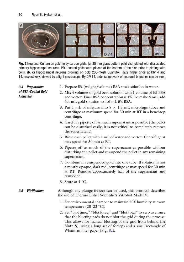
Fig. 2 Neuronal Culture on gold holey carbon grids. (a) 35 mm glass bottom petri dish plated with dissociated
primary hippocampal neurons. PDL-coated grids were placed at the bottom of the dish prior to plating with
cells. (b, c) Hippocampal neurons growing on gold 200-mesh Quantifoil R2/2 finder grids at DIV 4 and
14, respectively, viewed by a light microscope. By DIV 14, a dense network of neuronal branches can be seen
30
Ryan K. Hylton et al.
of BSA-Coated Gold
Fiducials
1. Prepare 5% (weight/volume) BSA stock solution in water.
2. Mix 4 volumes of gold bead solution with 1 volume of 5% BSA
and vortex. Final BSA concentration is 1%. To make 8 mL, add
6.4 mL gold solution to 1.6 mL 5% BSA.
3. Put 1 mL of mixture into 8 Â 1.5 mL microfuge tubes and
centrifuge at maximum speed for 30 min at RT in a benchtop
centrifuge.
4. Carefully pipette off as much supernatant as possible (the pellet
can be disturbed easily; it is not critical to completely remove
the supernatant).
5. Rinse each pellet with 1 mL of water and vortex. Centrifuge at
max speed for 30 min at RT.
6. Pipette off as much of the supernatant as possible without
disturbing the pellet and resuspend the pellet in any remaining
supernatant.
7. Combine all resuspended gold into one tube. If solution is not
a mostly opaque, dark red, centrifuge at max speed for 30 min
at RT. Remove approximately half of the supernatant and
resuspend.
8. Store at 4
C.
3.5 Vitrification
Although any plunge freezer can be used, this protocol describes
the use of Thermo Fisher Scientific’s Vitrobot Mark IV.
1. Set environmental chamber to maintain 70% humidity at room
temperature (20–22
C).
2. Set “blot time,” “blot force,” and “blot total” to zero to ensure
that the blotting pads do not blot the grid during the process.
This allows for manual blotting of the grid from behind (see
Note 8), using a long set of forceps and a small rectangle of
Whatman filter paper (Fig. 3a).
Fig. 2 Neuronal Culture on gold holey carbon grids. (a) 35 mm glass bottom petri dish plated with dissociated
primary hippocampal neurons. PDL-coated grids were placed at the bottom of the dish prior to plating with
cells. (b, c) Hippocampal neurons growing on gold 200-mesh Quantifoil R2/2 finder grids at DIV 4 and
14, respectively, viewed by a light microscope. By DIV 14, a dense network of neuronal branches can be seen
30
Ryan K. Hylton et al.