

In the last decade or so, tomographic data collection has
become semi-automated and a variety of software packages have
been developed for collecting image tilt-series in large batches. This
increased throughput has made it possible to routinely collect tens
of tomograms per day and upwards of 100 under ideal conditions.
Such a large amount of data is difficult to annotate and quantify, so
methods for automated tomogram segmentation have become a
necessity [5, 6]. In response, the software package EMAN2 was
updated in 2017 to include convolutional neural networks, which
can be trained to segment and annotate tomographic volumes [7].
Here, we detail and discuss a workflow for investigating the
ultrastructure of cultured hippocampal neurons using cryo-ET.
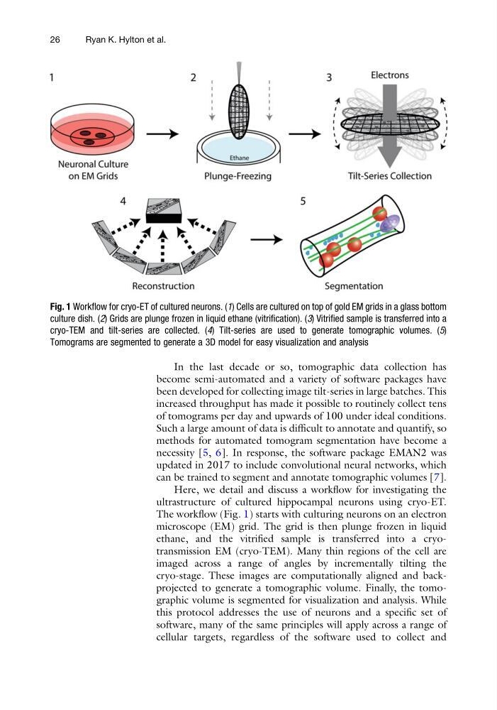
The workflow (Fig. 1) starts with culturing neurons on an electron
microscope (EM) grid. The grid is then plunge frozen in liquid
ethane, and the vitrified sample is transferred into a cryotransmission EM (cryo-TEM). Many thin regions of the cell are
imaged across a range of angles by incrementally tilting the
cryo-stage. These images are computationally aligned and backprojected to generate a tomographic volume. Finally, the tomographic volume is segmented for visualization and analysis. While
this protocol addresses the use of neurons and a specific set of
software, many of the same principles will apply across a range of
cellular targets, regardless of the software used to collect and
Fig. 1 Workflow for cryo-ET of cultured neurons. (1) Cells are cultured on top of gold EM grids in a glass bottom
culture dish. (2) Grids are plunge frozen in liquid ethane (vitrification). (3) Vitrified sample is transferred into a
cryo-TEM and tilt-series are collected. (4) Tilt-series are used to generate tomographic volumes. (5)
Tomograms are segmented to generate a 3D model for easy visualization and analysis
26
Ryan K. Hylton et al.
become semi-automated and a variety of software packages have
been developed for collecting image tilt-series in large batches. This
increased throughput has made it possible to routinely collect tens
of tomograms per day and upwards of 100 under ideal conditions.
Such a large amount of data is difficult to annotate and quantify, so
methods for automated tomogram segmentation have become a
necessity [5, 6]. In response, the software package EMAN2 was
updated in 2017 to include convolutional neural networks, which
can be trained to segment and annotate tomographic volumes [7].
Here, we detail and discuss a workflow for investigating the
ultrastructure of cultured hippocampal neurons using cryo-ET.
The workflow (Fig. 1) starts with culturing neurons on an electron
microscope (EM) grid. The grid is then plunge frozen in liquid
ethane, and the vitrified sample is transferred into a cryotransmission EM (cryo-TEM). Many thin regions of the cell are
imaged across a range of angles by incrementally tilting the
cryo-stage. These images are computationally aligned and backprojected to generate a tomographic volume. Finally, the tomographic volume is segmented for visualization and analysis. While
this protocol addresses the use of neurons and a specific set of
software, many of the same principles will apply across a range of
cellular targets, regardless of the software used to collect and
Fig. 1 Workflow for cryo-ET of cultured neurons. (1) Cells are cultured on top of gold EM grids in a glass bottom
culture dish. (2) Grids are plunge frozen in liquid ethane (vitrification). (3) Vitrified sample is transferred into a
cryo-TEM and tilt-series are collected. (4) Tilt-series are used to generate tomographic volumes. (5)
Tomograms are segmented to generate a 3D model for easy visualization and analysis
26
Ryan K. Hylton et al.