

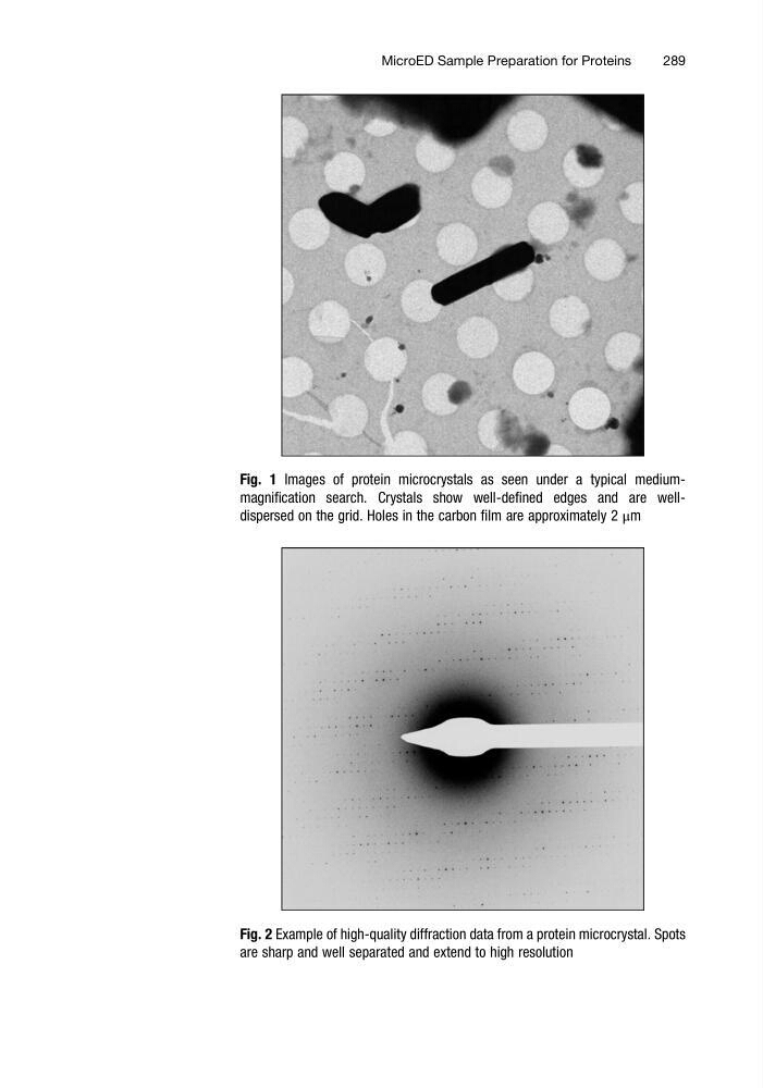
Fig. 1 Images of protein microcrystals as seen under a typical mediummagnification search. Crystals show well-defined edges and are welldispersed on the grid. Holes in the carbon film are approximately 2 μm
Fig. 2 Example of high-quality diffraction data from a protein microcrystal. Spots
are sharp and well separated and extend to high resolution
MicroED Sample Preparation for Proteins
289
Fig. 2 Example of high-quality diffraction data from a protein microcrystal. Spots
are sharp and well separated and extend to high resolution
MicroED Sample Preparation for Proteins
289