

3.2.4 Decision
on Cryo-EM Approach
Once crystals are identified, the crystal type will decide on the
approach for data collection and image processing. Single- and
double-layered 2D crystals, which tend to be the main goal of 2D
crystallization trials, are treated as described below (except for the
peel-blot in Subheading 3.3.2). These types of 2D crystals will have
sheet, vesicle, and/or planar-tubular morphology, not uncommonly in a mixture of morphologies. Narrow helical arrays of just
a few molecules across the diameter of the tube and typically several
hundred nanometers or more in lengths [19], will be ideal for the
helical analysis approach. Thin 3D crystals, usually consisting of 2D
crystals stacked in register, of approximately 1–2 μm and less than
1 μm thick will be tested for MicroED [10]. Smaller, stacked 2D
crystals, as well as crystals found not to be suitable for MicroED, are
subjected to the peel-blot approach.
3.3 Cryo-EM Grid
Preparation
and Cryo-EM
Single- or double-layered 2D crystals will be subjected to the steps
below, except for the peel-blot in Subheading 3.3.1, step 2. These
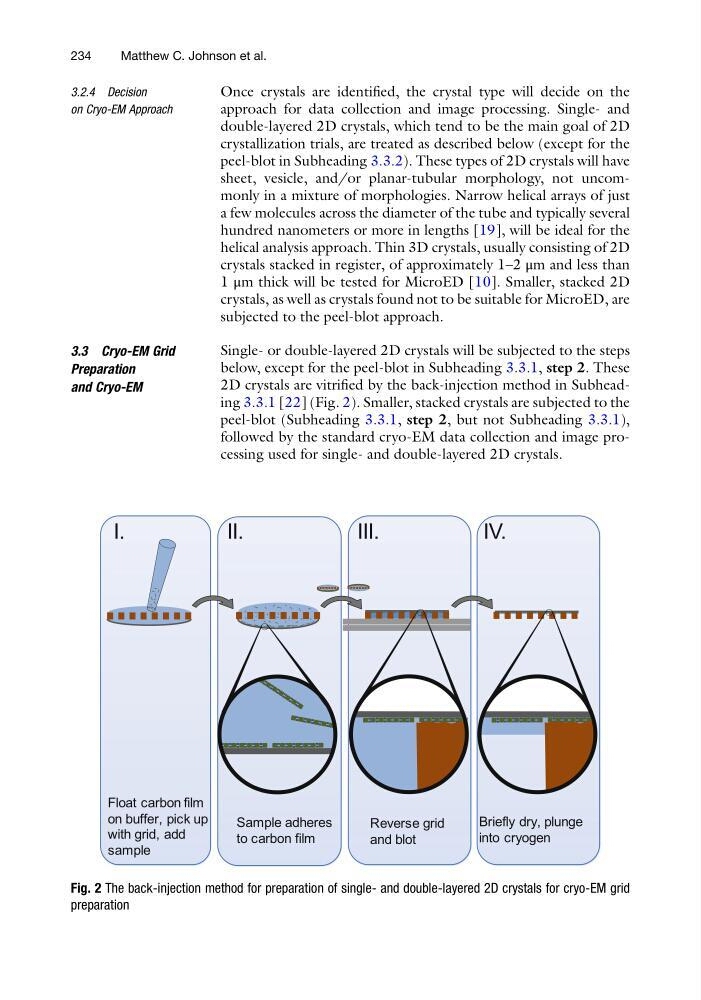
2D crystals are vitrified by the back-injection method in Subheading 3.3.1 [22] (Fig. 2). Smaller, stacked crystals are subjected to the
peel-blot (Subheading 3.3.1, step 2, but not Subheading 3.3.1),
followed by the standard cryo-EM data collection and image processing used for single- and double-layered 2D crystals.
I.
II.
III.
IV.
Sample adheres
to carbon film
Reverse grid
and blot
Briefly dry, plunge
into cryogen
Float carbon film
on buffer, pick up
with grid, add
sample
Fig. 2 The back-injection method for preparation of single- and double-layered 2D crystals for cryo-EM grid
preparation
234
Matthew C. Johnson et al.
on Cryo-EM Approach
Once crystals are identified, the crystal type will decide on the
approach for data collection and image processing. Single- and
double-layered 2D crystals, which tend to be the main goal of 2D
crystallization trials, are treated as described below (except for the
peel-blot in Subheading 3.3.2). These types of 2D crystals will have
sheet, vesicle, and/or planar-tubular morphology, not uncommonly in a mixture of morphologies. Narrow helical arrays of just
a few molecules across the diameter of the tube and typically several
hundred nanometers or more in lengths [19], will be ideal for the
helical analysis approach. Thin 3D crystals, usually consisting of 2D
crystals stacked in register, of approximately 1–2 μm and less than
1 μm thick will be tested for MicroED [10]. Smaller, stacked 2D
crystals, as well as crystals found not to be suitable for MicroED, are
subjected to the peel-blot approach.
3.3 Cryo-EM Grid
Preparation
and Cryo-EM
Single- or double-layered 2D crystals will be subjected to the steps
below, except for the peel-blot in Subheading 3.3.1, step 2. These
2D crystals are vitrified by the back-injection method in Subheading 3.3.1 [22] (Fig. 2). Smaller, stacked crystals are subjected to the
peel-blot (Subheading 3.3.1, step 2, but not Subheading 3.3.1),
followed by the standard cryo-EM data collection and image processing used for single- and double-layered 2D crystals.
I.
II.
III.
IV.
Sample adheres
to carbon film
Reverse grid
and blot
Briefly dry, plunge
into cryogen
Float carbon film
on buffer, pick up
with grid, add
sample
Fig. 2 The back-injection method for preparation of single- and double-layered 2D crystals for cryo-EM grid
preparation
234
Matthew C. Johnson et al.