

recent years has made it feasible to study decreasingly smaller
membrane proteins, which could previously only be studied via
crystallization. Nanodiscs [8] and styrene-maleic acid (SMA) nanodiscs [9] address the difficulties associated with studying purified
membrane proteins within a lipid bilayer rather than detergent,
which may cause issues for both the protein stability and cryo-EM
sample preparation. Yet many small proteins will remain outside the
reach of single particle cryo-EM, while neither 2D nor 3D electron
crystallography suffer from such a size limitation.
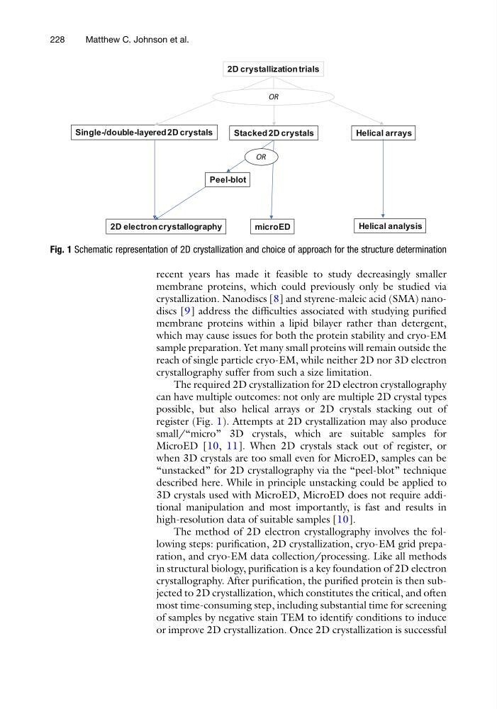
The required 2D crystallization for 2D electron crystallography
can have multiple outcomes: not only are multiple 2D crystal types
possible, but also helical arrays or 2D crystals stacking out of
register (Fig. 1). Attempts at 2D crystallization may also produce
small/“micro” 3D crystals, which are suitable samples for
MicroED [10, 11]. When 2D crystals stack out of register, or
when 3D crystals are too small even for MicroED, samples can be
“unstacked” for 2D crystallography via the “peel-blot” technique
described here. While in principle unstacking could be applied to
3D crystals used with MicroED, MicroED does not require additional manipulation and most importantly, is fast and results in
high-resolution data of suitable samples [10].
The method of 2D electron crystallography involves the following steps: purification, 2D crystallization, cryo-EM grid preparation, and cryo-EM data collection/processing. Like all methods
in structural biology, purification is a key foundation of 2D electron
crystallography. After purification, the purified protein is then subjected to 2D crystallization, which constitutes the critical, and often
most time-consuming step, including substantial time for screening
of samples by negative stain TEM to identify conditions to induce
or improve 2D crystallization. Once 2D crystallization is successful
Single-/double-layered 2D crystals
Stacked 2D crystals
2D electron crystallography
Helical arrays
Peel-blot
microED
Helical analysis
OR
2D crystallization trials
OR
Fig. 1 Schematic representation of 2D crystallization and choice of approach for the structure determination
228
Matthew C. Johnson et al.
membrane proteins, which could previously only be studied via
crystallization. Nanodiscs [8] and styrene-maleic acid (SMA) nanodiscs [9] address the difficulties associated with studying purified
membrane proteins within a lipid bilayer rather than detergent,
which may cause issues for both the protein stability and cryo-EM
sample preparation. Yet many small proteins will remain outside the
reach of single particle cryo-EM, while neither 2D nor 3D electron
crystallography suffer from such a size limitation.
The required 2D crystallization for 2D electron crystallography
can have multiple outcomes: not only are multiple 2D crystal types
possible, but also helical arrays or 2D crystals stacking out of
register (Fig. 1). Attempts at 2D crystallization may also produce
small/“micro” 3D crystals, which are suitable samples for
MicroED [10, 11]. When 2D crystals stack out of register, or
when 3D crystals are too small even for MicroED, samples can be
“unstacked” for 2D crystallography via the “peel-blot” technique
described here. While in principle unstacking could be applied to
3D crystals used with MicroED, MicroED does not require additional manipulation and most importantly, is fast and results in
high-resolution data of suitable samples [10].
The method of 2D electron crystallography involves the following steps: purification, 2D crystallization, cryo-EM grid preparation, and cryo-EM data collection/processing. Like all methods
in structural biology, purification is a key foundation of 2D electron
crystallography. After purification, the purified protein is then subjected to 2D crystallization, which constitutes the critical, and often
most time-consuming step, including substantial time for screening
of samples by negative stain TEM to identify conditions to induce
or improve 2D crystallization. Once 2D crystallization is successful
Single-/double-layered 2D crystals
Stacked 2D crystals
2D electron crystallography
Helical arrays
Peel-blot
microED
Helical analysis
OR
2D crystallization trials
OR
Fig. 1 Schematic representation of 2D crystallization and choice of approach for the structure determination
228
Matthew C. Johnson et al.