

focusing the diffraction pattern in the back focal plane of the
objective lens will ensure parallel illumination (Fig. 4).
1. Confirm that the specimen is at eucentric height and proper
focus at a magnification of SA 45,000Â in Nano probe mode.
Switch to Diffraction Mode (D 850 mm) and insert the
100 μm objective aperture if it is not already inserted. In the
Tecnai User Interface, click on the “Adjust” tab for the objective aperture and use the multifunction X and Y knobs to move
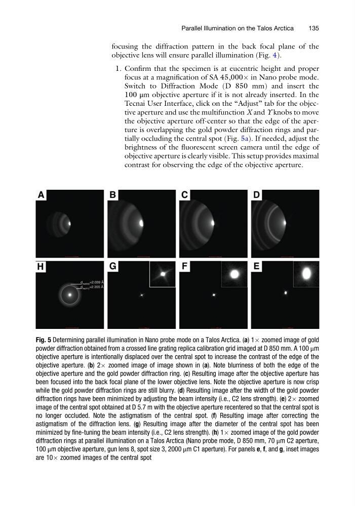
the objective aperture off-center so that the edge of the aperture is overlapping the gold powder diffraction rings and partially occluding the central spot (Fig. 5a). If needed, adjust the
brightness of the fluorescent screen camera until the edge of
objective aperture is clearly visible. This setup provides maximal
contrast for observing the edge of the objective aperture.
Fig. 5 Determining parallel illumination in Nano probe mode on a Talos Arctica. (a) 1Â zoomed image of gold
powder diffraction obtained from a crossed line grating replica calibration grid imaged at D 850 mm. A 100 μm
objective aperture is intentionally displaced over the central spot to increase the contrast of the edge of the
objective aperture. (b) 2Â zoomed image of image shown in (a). Note blurriness of both the edge of the
objective aperture and the gold powder diffraction ring. (c) Resulting image after the objective aperture has
been focused into the back focal plane of the lower objective lens. Note the objective aperture is now crisp
while the gold powder diffraction rings are still blurry. (d) Resulting image after the width of the gold powder
diffraction rings have been minimized by adjusting the beam intensity (i.e., C2 lens strength). (e) 2Â zoomed
image of the central spot obtained at D 5.7 m with the objective aperture recentered so that the central spot is
no longer occluded. Note the astigmatism of the central spot. (f) Resulting image after correcting the
astigmatism of the diffraction lens. (g) Resulting image after the diameter of the central spot has been
minimized by fine-tuning the beam intensity (i.e., C2 lens strength). (h) 1Â zoomed image of the gold powder
diffraction rings at parallel illumination on a Talos Arctica (Nano probe mode, D 850 mm, 70 μm C2 aperture,
100 μm objective aperture, gun lens 8, spot size 3, 2000 μm C1 aperture). For panels e, f, and g, inset images
are 10Â zoomed images of the central spot
Parallel Illumination on the Talos Arctica
135
objective lens will ensure parallel illumination (Fig. 4).
1. Confirm that the specimen is at eucentric height and proper
focus at a magnification of SA 45,000Â in Nano probe mode.
Switch to Diffraction Mode (D 850 mm) and insert the
100 μm objective aperture if it is not already inserted. In the
Tecnai User Interface, click on the “Adjust” tab for the objective aperture and use the multifunction X and Y knobs to move
the objective aperture off-center so that the edge of the aperture is overlapping the gold powder diffraction rings and partially occluding the central spot (Fig. 5a). If needed, adjust the
brightness of the fluorescent screen camera until the edge of
objective aperture is clearly visible. This setup provides maximal
contrast for observing the edge of the objective aperture.
Fig. 5 Determining parallel illumination in Nano probe mode on a Talos Arctica. (a) 1Â zoomed image of gold
powder diffraction obtained from a crossed line grating replica calibration grid imaged at D 850 mm. A 100 μm
objective aperture is intentionally displaced over the central spot to increase the contrast of the edge of the
objective aperture. (b) 2Â zoomed image of image shown in (a). Note blurriness of both the edge of the
objective aperture and the gold powder diffraction ring. (c) Resulting image after the objective aperture has
been focused into the back focal plane of the lower objective lens. Note the objective aperture is now crisp
while the gold powder diffraction rings are still blurry. (d) Resulting image after the width of the gold powder
diffraction rings have been minimized by adjusting the beam intensity (i.e., C2 lens strength). (e) 2Â zoomed
image of the central spot obtained at D 5.7 m with the objective aperture recentered so that the central spot is
no longer occluded. Note the astigmatism of the central spot. (f) Resulting image after correcting the
astigmatism of the diffraction lens. (g) Resulting image after the diameter of the central spot has been
minimized by fine-tuning the beam intensity (i.e., C2 lens strength). (h) 1Â zoomed image of the gold powder
diffraction rings at parallel illumination on a Talos Arctica (Nano probe mode, D 850 mm, 70 μm C2 aperture,
100 μm objective aperture, gun lens 8, spot size 3, 2000 μm C1 aperture). For panels e, f, and g, inset images
are 10Â zoomed images of the central spot
Parallel Illumination on the Talos Arctica
135