

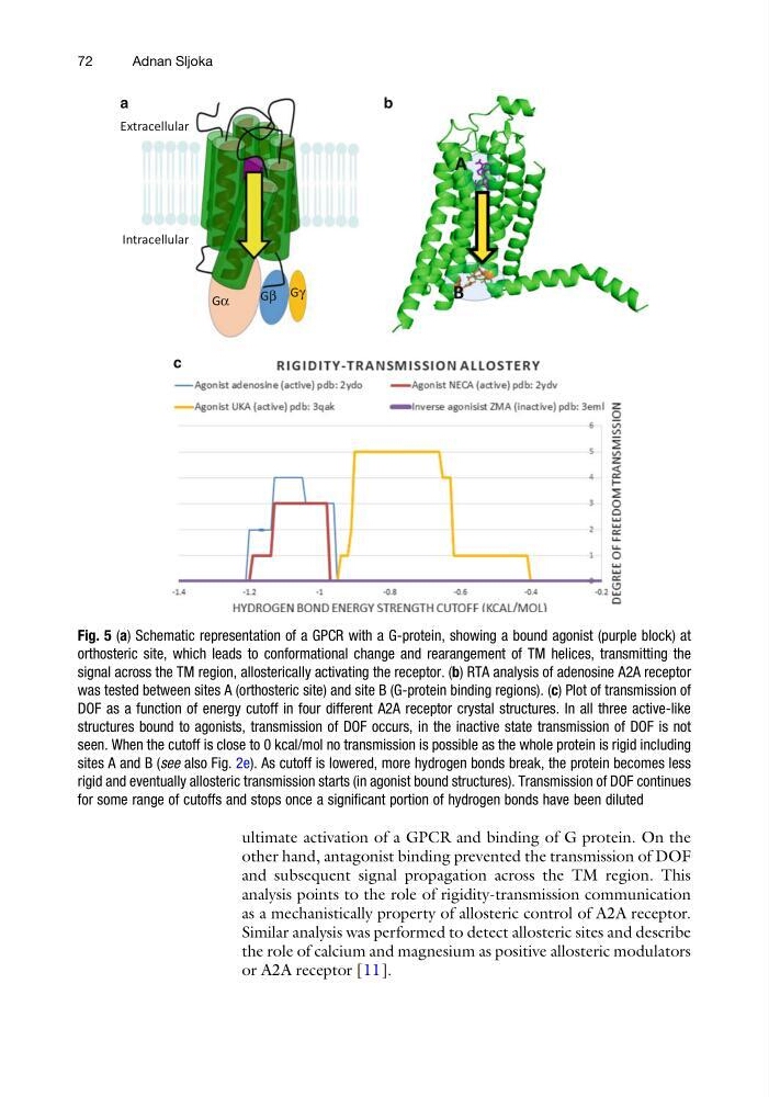
ultimate activation of a GPCR and binding of G protein. On the
other hand, antagonist binding prevented the transmission of DOF
and subsequent signal propagation across the TM region. This
analysis points to the role of rigidity-transmission communication
as a mechanistically property of allosteric control of A2A receptor.
Similar analysis was performed to detect allosteric sites and describe
the role of calcium and magnesium as positive allosteric modulators
or A2A receptor [11].
a
b
Gα
Gβ Gγ
Extracellular
Intracellular
A
B
c
Fig. 5 (a) Schematic representation of a GPCR with a G-protein, showing a bound agonist (purple block) at
orthosteric site, which leads to conformational change and rearangement of TM helices, transmitting the
signal across the TM region, allosterically activating the receptor. (b) RTA analysis of adenosine A2A receptor
was tested between sites A (orthosteric site) and site B (G-protein binding regions). (c) Plot of transmission of
DOF as a function of energy cutoff in four different A2A receptor crystal structures. In all three active-like
structures bound to agonists, transmission of DOF occurs, in the inactive state transmission of DOF is not
seen. When the cutoff is close to 0 kcal/mol no transmission is possible as the whole protein is rigid including
sites A and B (see also Fig. 2e). As cutoff is lowered, more hydrogen bonds break, the protein becomes less
rigid and eventually allosteric transmission starts (in agonist bound structures). Transmission of DOF continues
for some range of cutoffs and stops once a significant portion of hydrogen bonds have been diluted
72
Adnan Sljoka
other hand, antagonist binding prevented the transmission of DOF
and subsequent signal propagation across the TM region. This
analysis points to the role of rigidity-transmission communication
as a mechanistically property of allosteric control of A2A receptor.
Similar analysis was performed to detect allosteric sites and describe
the role of calcium and magnesium as positive allosteric modulators
or A2A receptor [11].
a
b
Gα
Gβ Gγ
Extracellular
Intracellular
A
B
c
Fig. 5 (a) Schematic representation of a GPCR with a G-protein, showing a bound agonist (purple block) at
orthosteric site, which leads to conformational change and rearangement of TM helices, transmitting the
signal across the TM region, allosterically activating the receptor. (b) RTA analysis of adenosine A2A receptor
was tested between sites A (orthosteric site) and site B (G-protein binding regions). (c) Plot of transmission of
DOF as a function of energy cutoff in four different A2A receptor crystal structures. In all three active-like
structures bound to agonists, transmission of DOF occurs, in the inactive state transmission of DOF is not
seen. When the cutoff is close to 0 kcal/mol no transmission is possible as the whole protein is rigid including
sites A and B (see also Fig. 2e). As cutoff is lowered, more hydrogen bonds break, the protein becomes less
rigid and eventually allosteric transmission starts (in agonist bound structures). Transmission of DOF continues
for some range of cutoffs and stops once a significant portion of hydrogen bonds have been diluted
72
Adnan Sljoka