

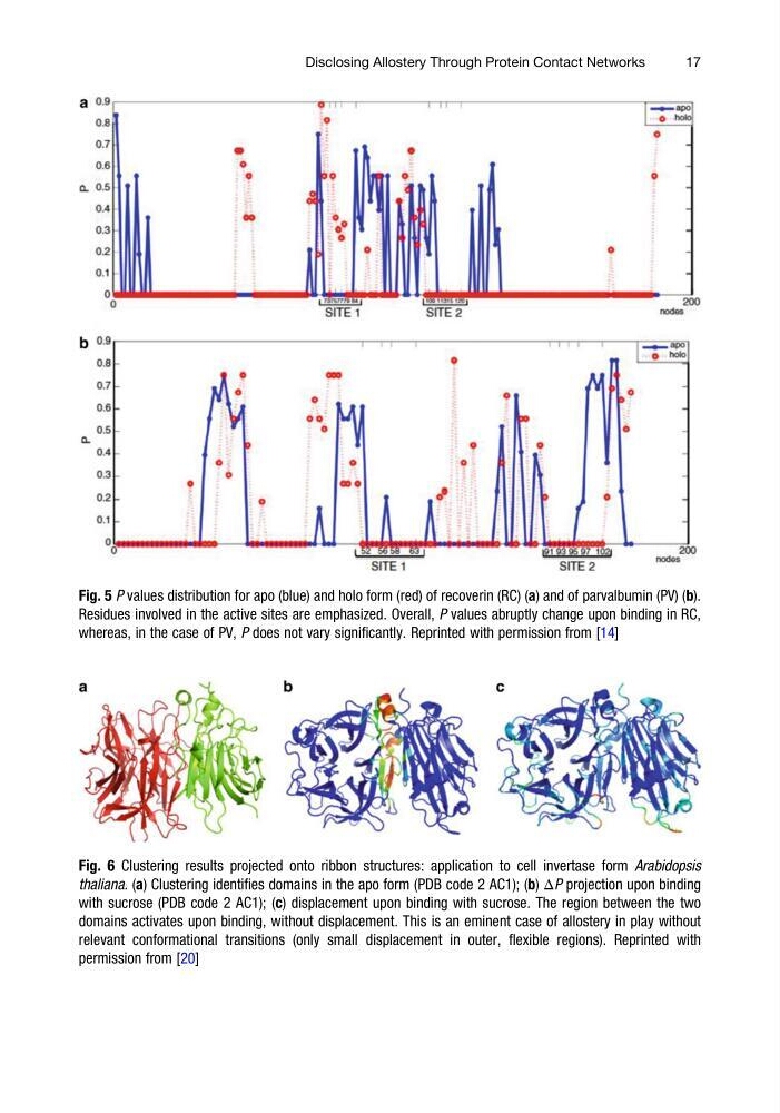
Fig. 6 Clustering results projected onto ribbon structures: application to cell invertase form Arabidopsis
thaliana. (a) Clustering identifies domains in the apo form (PDB code 2 AC1); (b) ΔP projection upon binding
with sucrose (PDB code 2 AC1); (c) displacement upon binding with sucrose. The region between the two
domains activates upon binding, without displacement. This is an eminent case of allostery in play without
relevant conformational transitions (only small displacement in outer, flexible regions). Reprinted with
permission from [20]
Fig. 5 P values distribution for apo (blue) and holo form (red) of recoverin (RC) (a) and of parvalbumin (PV) (b).
Residues involved in the active sites are emphasized. Overall, P values abruptly change upon binding in RC,
whereas, in the case of PV, P does not vary significantly. Reprinted with permission from [14]
Disclosing Allostery Through Protein Contact Networks
17
thaliana. (a) Clustering identifies domains in the apo form (PDB code 2 AC1); (b) ΔP projection upon binding
with sucrose (PDB code 2 AC1); (c) displacement upon binding with sucrose. The region between the two
domains activates upon binding, without displacement. This is an eminent case of allostery in play without
relevant conformational transitions (only small displacement in outer, flexible regions). Reprinted with
permission from [20]
Fig. 5 P values distribution for apo (blue) and holo form (red) of recoverin (RC) (a) and of parvalbumin (PV) (b).
Residues involved in the active sites are emphasized. Overall, P values abruptly change upon binding in RC,
whereas, in the case of PV, P does not vary significantly. Reprinted with permission from [14]
Disclosing Allostery Through Protein Contact Networks
17