

S148 has also been shown to play an important role in the
occluded conformation of ecDHFR through mutational analysis.
The occluded conformation is stabilized via two hydrogen bonds
between Asn23 in the Met20 loop and Ser148. In a study conducted by Behiry et al. S148 was replaced with proline (S148P).
Pro cannot form the hydrogen bond interactions necessary to
stabilize the occluded conformation and the S148P mutant is
useful for investigating the importance of the occluded conformation in DHFR catalysis. Their results indicated that the occluded
conformation assists in the release of the oxidized cofactor NADP
+
and progression through the catalytic cycle [97].
5.5 Mutations
in the DHF
Binding Cleft
The residues in the DHF binding site of ecDHFR are I5, A6, A7,
M20, D27, L28, F31, R52, R57, I94, and T113. Among these
residues, the backbone of I5 and side chains of D27, R52, R57
directly interact with DHF via hydrogen bonding interactions
[98]. The other residues can be considered distal in this context
because they are not directly involved in binding of substrate or the
catalytic reaction. In a recent study by Abdizadeh et al., replacement of L28 with an Arg residue (L28R) was investigated to
structurally and dynamically characterize the WT and L28R
ecDHFR in the presence of DHF and TMP. They used the
NAMD package to model the molecular dynamics of the protein–
water systems to determine the conformational space, loop dynamics, and hydrogen binding interactions for the WT and mutant
enzyme. The protein was soaked in a cubic solvent box with at
least a 10 A ˚ layer of solvent in each direction from any atom of the
protein to the edge of the box and the ionic strength in the
simulations was kept at 150 mM. All systems were subjected to
10,000 steps of energy minimization. The resulting structures were
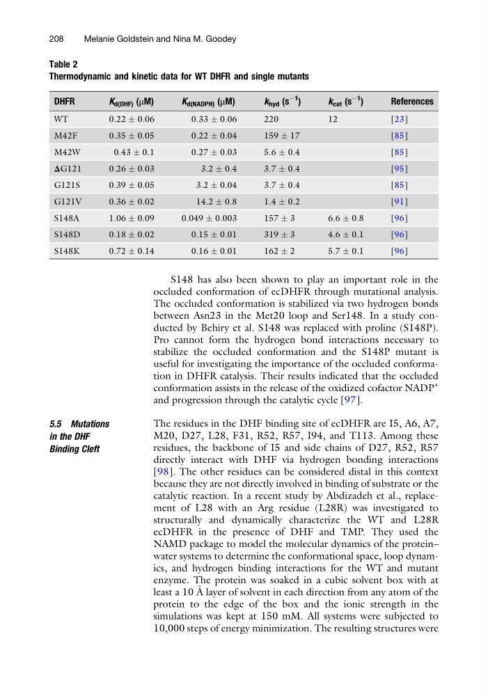
Table 2
Thermodynamic and kinetic data for WT DHFR and single mutants
DHFR
K d(DHF) (μM)
K d(NADPH) (μM)
k hyd (s
À1
)
k cat (s
À1
)
References
WT
0.22 Æ 0.06
0.33 Æ 0.06
220
12
[23]
M42F
0.35 Æ 0.05
0.22 Æ 0.04
159 Æ 17
[85]
M42W
0.43 Æ 0.1
0.27 Æ 0.03
5.6 Æ 0.4
[85]
ΔG121
0.26 Æ 0.03
3.2 Æ 0.4
3.7 Æ 0.4
[95]
G121S
0.39 Æ 0.05
3.2 Æ 0.04
3.7 Æ 0.4
[85]
G121V
0.36 Æ 0.02
14.2 Æ 0.8
1.4 Æ 0.2
[91]
S148A
1.06 Æ 0.09
0.049 Æ 0.003
157 Æ 3
6.6 Æ 0.8
[96]
S148D
0.18 Æ 0.02
0.15 Æ 0.01
319 Æ 3
4.6 Æ 0.1
[96]
S148K
0.72 Æ 0.14
0.16 Æ 0.01
162 Æ 2
5.7 Æ 0.1
[96]
208
Melanie Goldstein and Nina M. Goodey
occluded conformation of ecDHFR through mutational analysis.
The occluded conformation is stabilized via two hydrogen bonds
between Asn23 in the Met20 loop and Ser148. In a study conducted by Behiry et al. S148 was replaced with proline (S148P).
Pro cannot form the hydrogen bond interactions necessary to
stabilize the occluded conformation and the S148P mutant is
useful for investigating the importance of the occluded conformation in DHFR catalysis. Their results indicated that the occluded
conformation assists in the release of the oxidized cofactor NADP
+
and progression through the catalytic cycle [97].
5.5 Mutations
in the DHF
Binding Cleft
The residues in the DHF binding site of ecDHFR are I5, A6, A7,
M20, D27, L28, F31, R52, R57, I94, and T113. Among these
residues, the backbone of I5 and side chains of D27, R52, R57
directly interact with DHF via hydrogen bonding interactions
[98]. The other residues can be considered distal in this context
because they are not directly involved in binding of substrate or the
catalytic reaction. In a recent study by Abdizadeh et al., replacement of L28 with an Arg residue (L28R) was investigated to
structurally and dynamically characterize the WT and L28R
ecDHFR in the presence of DHF and TMP. They used the
NAMD package to model the molecular dynamics of the protein–
water systems to determine the conformational space, loop dynamics, and hydrogen binding interactions for the WT and mutant
enzyme. The protein was soaked in a cubic solvent box with at
least a 10 A ˚ layer of solvent in each direction from any atom of the
protein to the edge of the box and the ionic strength in the
simulations was kept at 150 mM. All systems were subjected to
10,000 steps of energy minimization. The resulting structures were
Table 2
Thermodynamic and kinetic data for WT DHFR and single mutants
DHFR
K d(DHF) (μM)
K d(NADPH) (μM)
k hyd (s
À1
)
k cat (s
À1
)
References
WT
0.22 Æ 0.06
0.33 Æ 0.06
220
12
[23]
M42F
0.35 Æ 0.05
0.22 Æ 0.04
159 Æ 17
[85]
M42W
0.43 Æ 0.1
0.27 Æ 0.03
5.6 Æ 0.4
[85]
ΔG121
0.26 Æ 0.03
3.2 Æ 0.4
3.7 Æ 0.4
[95]
G121S
0.39 Æ 0.05
3.2 Æ 0.04
3.7 Æ 0.4
[85]
G121V
0.36 Æ 0.02
14.2 Æ 0.8
1.4 Æ 0.2
[91]
S148A
1.06 Æ 0.09
0.049 Æ 0.003
157 Æ 3
6.6 Æ 0.8
[96]
S148D
0.18 Æ 0.02
0.15 Æ 0.01
319 Æ 3
4.6 Æ 0.1
[96]
S148K
0.72 Æ 0.14
0.16 Æ 0.01
162 Æ 2
5.7 Æ 0.1
[96]
208
Melanie Goldstein and Nina M. Goodey