

5.1 Mutations
in the Adenosine
Binding Subdomain
M42 in ecDHFR is a highly conserved residue located in the
hydrophobic core of the adenosine binding domain, approximately
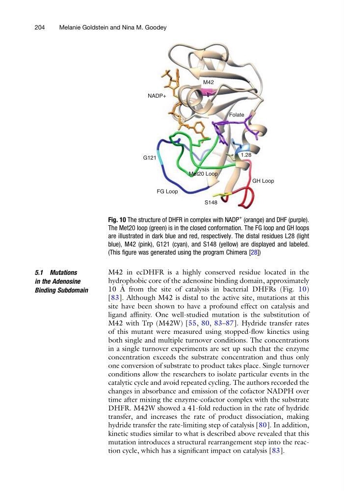
10 A ˚ from the site of catalysis in bacterial DHFRs (Fig. 10)
[83]. Although M42 is distal to the active site, mutations at this
site have been shown to have a profound effect on catalysis and
ligand affinity. One well-studied mutation is the substitution of
M42 with Trp (M42W) [55, 80, 83–87]. Hydride transfer rates
of this mutant were measured using stopped-flow kinetics using
both single and multiple turnover conditions. The concentrations
in a single turnover experiments are set up such that the enzyme
concentration exceeds the substrate concentration and thus only
one conversion of substrate to product takes place. Single turnover
conditions allow the researchers to isolate particular events in the
catalytic cycle and avoid repeated cycling. The authors recorded the
changes in absorbance and emission of the cofactor NADPH over
time after mixing the enzyme-cofactor complex with the substrate
DHFR. M42W showed a 41-fold reduction in the rate of hydride
transfer, and increases the rate of product dissociation, making
hydride transfer the rate-limiting step of catalysis [80]. In addition,
kinetic studies similar to what is described above revealed that this
mutation introduces a structural rearrangement step into the reaction cycle, which has a significant impact on catalysis [83].
GH Loop
1.28
Folate
S148
Met20 Loop
NADP+
M42
FG Loop
G121
Fig. 10 The structure of DHFR in complex with NADP
+
(orange) and DHF (purple).
The Met20 loop (green) is in the closed conformation. The FG loop and GH loops
are illustrated in dark blue and red, respectively. The distal residues L28 (light
blue), M42 (pink), G121 (cyan), and S148 (yellow) are displayed and labeled.
(This figure was generated using the program Chimera [28])
204
Melanie Goldstein and Nina M. Goodey
in the Adenosine
Binding Subdomain
M42 in ecDHFR is a highly conserved residue located in the
hydrophobic core of the adenosine binding domain, approximately
10 A ˚ from the site of catalysis in bacterial DHFRs (Fig. 10)
[83]. Although M42 is distal to the active site, mutations at this
site have been shown to have a profound effect on catalysis and
ligand affinity. One well-studied mutation is the substitution of
M42 with Trp (M42W) [55, 80, 83–87]. Hydride transfer rates
of this mutant were measured using stopped-flow kinetics using
both single and multiple turnover conditions. The concentrations
in a single turnover experiments are set up such that the enzyme
concentration exceeds the substrate concentration and thus only
one conversion of substrate to product takes place. Single turnover
conditions allow the researchers to isolate particular events in the
catalytic cycle and avoid repeated cycling. The authors recorded the
changes in absorbance and emission of the cofactor NADPH over
time after mixing the enzyme-cofactor complex with the substrate
DHFR. M42W showed a 41-fold reduction in the rate of hydride
transfer, and increases the rate of product dissociation, making
hydride transfer the rate-limiting step of catalysis [80]. In addition,
kinetic studies similar to what is described above revealed that this
mutation introduces a structural rearrangement step into the reaction cycle, which has a significant impact on catalysis [83].
GH Loop
1.28
Folate
S148
Met20 Loop
NADP+
M42
FG Loop
G121
Fig. 10 The structure of DHFR in complex with NADP
+
(orange) and DHF (purple).
The Met20 loop (green) is in the closed conformation. The FG loop and GH loops
are illustrated in dark blue and red, respectively. The distal residues L28 (light
blue), M42 (pink), G121 (cyan), and S148 (yellow) are displayed and labeled.
(This figure was generated using the program Chimera [28])
204
Melanie Goldstein and Nina M. Goodey