

These loop regions serve as a gate, closing over the bound ligands
in the ternary E:DHF:NADPH complex, but are mobile in the
apoenzyme and holoenzyme [27]. The substrate and cofactor
bind in a hydrophobic cleft at the juncture of the two subdomains
and hinge bending motions about residues Lys38 and Val88 allow
the adenosine binding domain to move relative to the major
domain upon ligand binding, closing off the active site cleft to the
surrounding environment (Fig. 2) [18, 27].
2.2 Active Site
Structure and Loop
Conformations
In the ternary complex, the pterin ring of the substrate and the
nicotinamide ring of the cofactor bind in close proximity in the
active site, with the hydride donor atom (C4 of NADPH) and the
hydride acceptor (C6 of the pterin ring) in van der Waals contact
[29]. The DHFR active site contains an invariant carboxylic acid
residue, Asp27 in bacteria and a glutamic acid residue in vertebrates
(Glu30 in humans) [30]. Mutational studies have supported that
Asp27 has a crucial role in the hydride transfer step [30, 31].
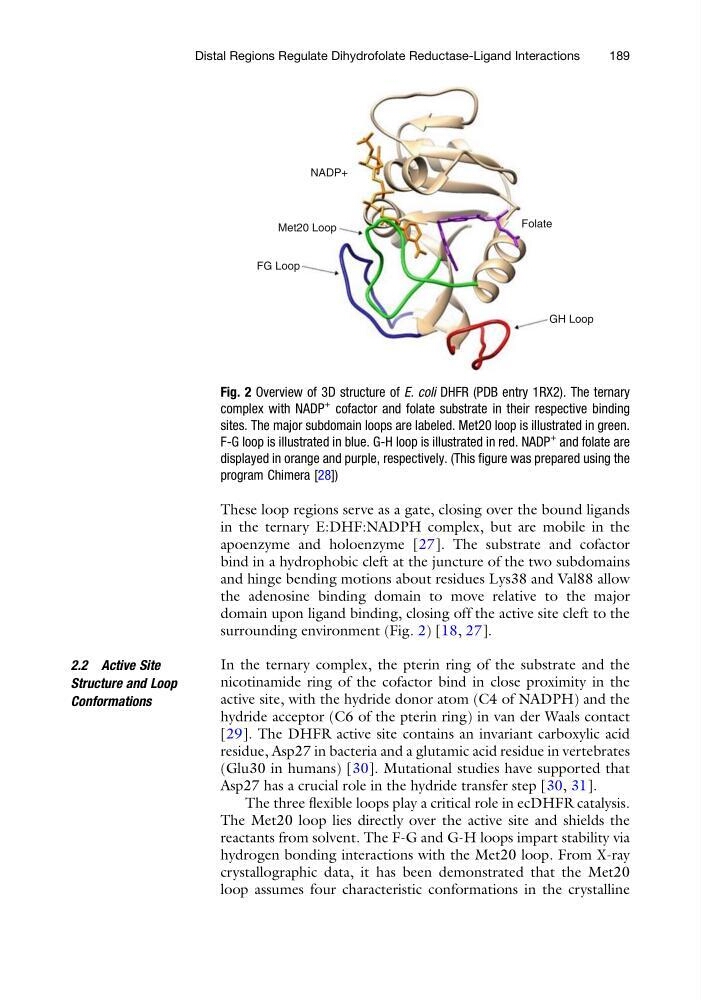
The three flexible loops play a critical role in ecDHFR catalysis.
The Met20 loop lies directly over the active site and shields the
reactants from solvent. The F-G and G-H loops impart stability via
hydrogen bonding interactions with the Met20 loop. From X-ray
crystallographic data, it has been demonstrated that the Met20
loop assumes four characteristic conformations in the crystalline
NADP+
Met20 Loop
FG Loop
GH Loop
Folate
Fig. 2 Overview of 3D structure of E. coli DHFR (PDB entry 1RX2). The ternary
complex with NADP
+
cofactor and folate substrate in their respective binding
sites. The major subdomain loops are labeled. Met20 loop is illustrated in green.
F-G loop is illustrated in blue. G-H loop is illustrated in red. NADP
+
and folate are
displayed in orange and purple, respectively. (This figure was prepared using the
program Chimera [28])
Distal Regions Regulate Dihydrofolate Reductase-Ligand Interactions
189
in the ternary E:DHF:NADPH complex, but are mobile in the
apoenzyme and holoenzyme [27]. The substrate and cofactor
bind in a hydrophobic cleft at the juncture of the two subdomains
and hinge bending motions about residues Lys38 and Val88 allow
the adenosine binding domain to move relative to the major
domain upon ligand binding, closing off the active site cleft to the
surrounding environment (Fig. 2) [18, 27].
2.2 Active Site
Structure and Loop
Conformations
In the ternary complex, the pterin ring of the substrate and the
nicotinamide ring of the cofactor bind in close proximity in the
active site, with the hydride donor atom (C4 of NADPH) and the
hydride acceptor (C6 of the pterin ring) in van der Waals contact
[29]. The DHFR active site contains an invariant carboxylic acid
residue, Asp27 in bacteria and a glutamic acid residue in vertebrates
(Glu30 in humans) [30]. Mutational studies have supported that
Asp27 has a crucial role in the hydride transfer step [30, 31].
The three flexible loops play a critical role in ecDHFR catalysis.
The Met20 loop lies directly over the active site and shields the
reactants from solvent. The F-G and G-H loops impart stability via
hydrogen bonding interactions with the Met20 loop. From X-ray
crystallographic data, it has been demonstrated that the Met20
loop assumes four characteristic conformations in the crystalline
NADP+
Met20 Loop
FG Loop
GH Loop
Folate
Fig. 2 Overview of 3D structure of E. coli DHFR (PDB entry 1RX2). The ternary
complex with NADP
+
cofactor and folate substrate in their respective binding
sites. The major subdomain loops are labeled. Met20 loop is illustrated in green.
F-G loop is illustrated in blue. G-H loop is illustrated in red. NADP
+
and folate are
displayed in orange and purple, respectively. (This figure was prepared using the
program Chimera [28])
Distal Regions Regulate Dihydrofolate Reductase-Ligand Interactions
189