

3. A 500-step steepest descent (SD) minimization with backbone
atoms fixed is used to relax the initial structures of antibody and
prion peptide.
2.1.2 Structures
of Antibody-Antigen
Complexes
1. The structures of the bound forms of the Fab/peptide complex
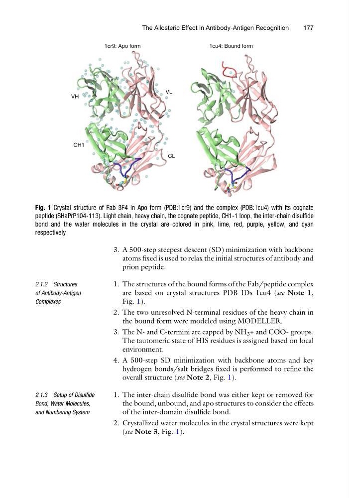
are based on crystal structures PDB IDs 1cu4 (see Note 1,
Fig. 1).
2. The two unresolved N-terminal residues of the heavy chain in
the bound form were modeled using MODELLER.
3. The N- and C-termini are capped by NH 3 + and COO- groups.
The tautomeric state of HIS residues is assigned based on local
environment.
4. A 500-step SD minimization with backbone atoms and key
hydrogen bonds/salt bridges fixed is performed to refine the
overall structure (see Note 2, Fig. 1).
2.1.3 Setup of Disulfide
Bond, Water Molecules,
and Numbering System
1. The inter-chain disulfide bond was either kept or removed for
the bound, unbound, and apo structures to consider the effects
of the inter-domain disulfide bond.
2. Crystallized water molecules in the crystal structures were kept
(see Note 3, Fig. 1).
VH
VL
CL
1cr9: Apo form
CH1
1cu4: Bound form
Fig. 1 Crystal structure of Fab 3F4 in Apo form (PDB:1cr9) and the complex (PDB:1cu4) with its cognate
peptide (SHaPrP104-113). Light chain, heavy chain, the cognate peptide, CH1-1 loop, the inter-chain disulfide
bond and the water molecules in the crystal are colored in pink, lime, red, purple, yellow, and cyan
respectively
The Allosteric Effect in Antibody-Antigen Recognition
177
atoms fixed is used to relax the initial structures of antibody and
prion peptide.
2.1.2 Structures
of Antibody-Antigen
Complexes
1. The structures of the bound forms of the Fab/peptide complex
are based on crystal structures PDB IDs 1cu4 (see Note 1,
Fig. 1).
2. The two unresolved N-terminal residues of the heavy chain in
the bound form were modeled using MODELLER.
3. The N- and C-termini are capped by NH 3 + and COO- groups.
The tautomeric state of HIS residues is assigned based on local
environment.
4. A 500-step SD minimization with backbone atoms and key
hydrogen bonds/salt bridges fixed is performed to refine the
overall structure (see Note 2, Fig. 1).
2.1.3 Setup of Disulfide
Bond, Water Molecules,
and Numbering System
1. The inter-chain disulfide bond was either kept or removed for
the bound, unbound, and apo structures to consider the effects
of the inter-domain disulfide bond.
2. Crystallized water molecules in the crystal structures were kept
(see Note 3, Fig. 1).
VH
VL
CL
1cr9: Apo form
CH1
1cu4: Bound form
Fig. 1 Crystal structure of Fab 3F4 in Apo form (PDB:1cr9) and the complex (PDB:1cu4) with its cognate
peptide (SHaPrP104-113). Light chain, heavy chain, the cognate peptide, CH1-1 loop, the inter-chain disulfide
bond and the water molecules in the crystal are colored in pink, lime, red, purple, yellow, and cyan
respectively
The Allosteric Effect in Antibody-Antigen Recognition
177