

4
of metals by the microorganisms from the natural substrate is achieved by various
strategies such as cleavage of chemical bonds, chelation, siderophore-mediated iron
acquisition, and redox reactions.
1.3 Classification of Fungal Siderophores
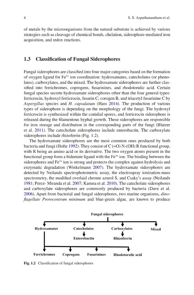
Fungal siderophores are classified into four major categories based on the formation
of oxygen ligand for Fe
3+
ion coordination: hydroxamates, catecholates (or phenolates), carboxylates, and the mixed. The hydroxamate siderophores are further classified into ferrichromes, coprogens, fusarinines, and rhodotorulic acid. Certain
fungal species secrete hydroxamate siderophores other than the four general types:
ferricrocin, hydroxyl ferricrocin, fusarin C, corogen B, and triacetyl fusarinine C by
Aspergillus species and H. capsulatum (Hass 2014). The production of various
types of siderophore is depending on the morphology of the fungi. The hydroxyl
ferricrocin is synthesized within the conidial spores, and ferricrocin siderophore is
released during the filamentous hyphal growth. These siderophores are responsible
for iron storage and distribution in the corresponding parts of the fungi (Blatzer
et al. 2011). The catecholate siderophores include enterobactin. The carboxylate
siderophores include rhizoferrin (Fig. 1.2).
The hydroxamate siderophores are the most common ones produced by both
bacteria and fungi (Hofte 1992). They consist of C (=O) N-(OH) R functional group,
with R being an amino acid or its derivative. The two oxygen atoms present in the
functional group form a bidentate ligand with the Fe
3+
ion. The binding between the
siderophores and Fe
3+
ion is strong and protects the complex against hydrolysis and
enzymatic degradation (Winkelmann 2007). The hydroxamate siderophores are
detected by Neilands spectrophotometric assay, the electrospray ionization-mass
spectrometry, the modified overlaid chrome azurol S, and Csaky’s assay (Neilands
1981; Perez- Miranda et al. 2007; Karuna et al. 2010). The catecholate siderophores
and carboxylate siderophores are commonly produced by bacteria (Dave et al.
2006). Apart from bacterial and fungal siderophores, two marine organisms, dinoflagellate Prorocentrum minimum and blue-green algae, are known to produce
Fig. 1.2 Classification of fungal siderophores
S. S. Arputhanantham et al.
of metals by the microorganisms from the natural substrate is achieved by various
strategies such as cleavage of chemical bonds, chelation, siderophore-mediated iron
acquisition, and redox reactions.
1.3 Classification of Fungal Siderophores
Fungal siderophores are classified into four major categories based on the formation
of oxygen ligand for Fe
3+
ion coordination: hydroxamates, catecholates (or phenolates), carboxylates, and the mixed. The hydroxamate siderophores are further classified into ferrichromes, coprogens, fusarinines, and rhodotorulic acid. Certain
fungal species secrete hydroxamate siderophores other than the four general types:
ferricrocin, hydroxyl ferricrocin, fusarin C, corogen B, and triacetyl fusarinine C by
Aspergillus species and H. capsulatum (Hass 2014). The production of various
types of siderophore is depending on the morphology of the fungi. The hydroxyl
ferricrocin is synthesized within the conidial spores, and ferricrocin siderophore is
released during the filamentous hyphal growth. These siderophores are responsible
for iron storage and distribution in the corresponding parts of the fungi (Blatzer
et al. 2011). The catecholate siderophores include enterobactin. The carboxylate
siderophores include rhizoferrin (Fig. 1.2).
The hydroxamate siderophores are the most common ones produced by both
bacteria and fungi (Hofte 1992). They consist of C (=O) N-(OH) R functional group,
with R being an amino acid or its derivative. The two oxygen atoms present in the
functional group form a bidentate ligand with the Fe
3+
ion. The binding between the
siderophores and Fe
3+
ion is strong and protects the complex against hydrolysis and
enzymatic degradation (Winkelmann 2007). The hydroxamate siderophores are
detected by Neilands spectrophotometric assay, the electrospray ionization-mass
spectrometry, the modified overlaid chrome azurol S, and Csaky’s assay (Neilands
1981; Perez- Miranda et al. 2007; Karuna et al. 2010). The catecholate siderophores
and carboxylate siderophores are commonly produced by bacteria (Dave et al.
2006). Apart from bacterial and fungal siderophores, two marine organisms, dinoflagellate Prorocentrum minimum and blue-green algae, are known to produce
Fig. 1.2 Classification of fungal siderophores
S. S. Arputhanantham et al.