

356
14.6 Physical Processes Underlying Leaf Optical Properties
Leaf anatomy and phylogeny determine how leaf cells (epidermis, mesophyll) and
specialized tissues (xylem and phloem conducting tissues and the stomatal complex) are arranged (Al-Edany and Al-Saadi 2012) and how this affects reflectance.
There are many modifications, but the standard arrangement of cells and tissues in
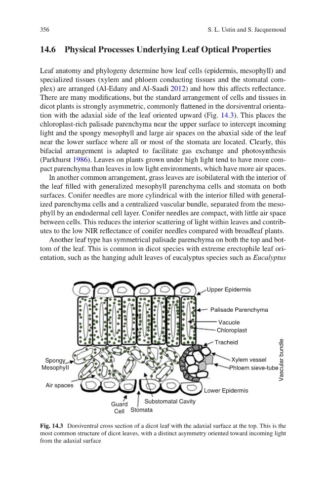
dicot plants is strongly asymmetric, commonly flattened in the dorsiventral orientation with the adaxial side of the leaf oriented upward (Fig. 14.3). This places the
chloroplast-rich palisade parenchyma near the upper surface to intercept incoming
light and the spongy mesophyll and large air spaces on the abaxial side of the leaf
near the lower surface where all or most of the stomata are located. Clearly, this
bifacial arrangement is adapted to facilitate gas exchange and photosynthesis
(Parkhurst 1986). Leaves on plants grown under high light tend to have more compact parenchyma than leaves in low light environments, which have more air spaces.
In another common arrangement, grass leaves are isobilateral with the interior of
the leaf filled with generalized mesophyll parenchyma cells and stomata on both
surfaces. Conifer needles are more cylindrical with the interior filled with generalized parenchyma cells and a centralized vascular bundle, separated from the mesophyll by an endodermal cell layer. Conifer needles are compact, with little air space
between cells. This reduces the interior scattering of light within leaves and contributes to the low NIR reflectance of conifer needles compared with broadleaf plants.
Another leaf type has symmetrical palisade parenchyma on both the top and bottom of the leaf. This is common in dicot species with extreme erectophile leaf orientation, such as the hanging adult leaves of eucalyptus species such as Eucalyptus
Substomatal Cavity
Stomata
Guard
Cell
Air spaces
Spongy
MesophyII
Lower Epidermis
Tracheid
Palisade Parenchyma
Upper Epidermis
Chloroplast
Vacuole
Vascular bundle
Xylem vessel
Phloem sieve-tube
Fig. 14.3 Dorsiventral cross section of a dicot leaf with the adaxial surface at the top. This is the
most common structure of dicot leaves, with a distinct asymmetry oriented toward incoming light
from the adaxial surface
S. L. Ustin and S. Jacquemoud
14.6 Physical Processes Underlying Leaf Optical Properties
Leaf anatomy and phylogeny determine how leaf cells (epidermis, mesophyll) and
specialized tissues (xylem and phloem conducting tissues and the stomatal complex) are arranged (Al-Edany and Al-Saadi 2012) and how this affects reflectance.
There are many modifications, but the standard arrangement of cells and tissues in
dicot plants is strongly asymmetric, commonly flattened in the dorsiventral orientation with the adaxial side of the leaf oriented upward (Fig. 14.3). This places the
chloroplast-rich palisade parenchyma near the upper surface to intercept incoming
light and the spongy mesophyll and large air spaces on the abaxial side of the leaf
near the lower surface where all or most of the stomata are located. Clearly, this
bifacial arrangement is adapted to facilitate gas exchange and photosynthesis
(Parkhurst 1986). Leaves on plants grown under high light tend to have more compact parenchyma than leaves in low light environments, which have more air spaces.
In another common arrangement, grass leaves are isobilateral with the interior of
the leaf filled with generalized mesophyll parenchyma cells and stomata on both
surfaces. Conifer needles are more cylindrical with the interior filled with generalized parenchyma cells and a centralized vascular bundle, separated from the mesophyll by an endodermal cell layer. Conifer needles are compact, with little air space
between cells. This reduces the interior scattering of light within leaves and contributes to the low NIR reflectance of conifer needles compared with broadleaf plants.
Another leaf type has symmetrical palisade parenchyma on both the top and bottom of the leaf. This is common in dicot species with extreme erectophile leaf orientation, such as the hanging adult leaves of eucalyptus species such as Eucalyptus
Substomatal Cavity
Stomata
Guard
Cell
Air spaces
Spongy
MesophyII
Lower Epidermis
Tracheid
Palisade Parenchyma
Upper Epidermis
Chloroplast
Vacuole
Vascular bundle
Xylem vessel
Phloem sieve-tube
Fig. 14.3 Dorsiventral cross section of a dicot leaf with the adaxial surface at the top. This is the
most common structure of dicot leaves, with a distinct asymmetry oriented toward incoming light
from the adaxial surface
S. L. Ustin and S. Jacquemoud