

fiducial markers for single-particle cryo-electron microscopy (cryoEM) to aid in structure determination [8, 9].
The preparation of milligram quantities of purified Fv and Fab
fragments is the most critical prerequisite once the strategies for
antibody-aided structural studies are adopted. Recently, we developed an efficient production strategy for recombinant Fv antibody
fragments and named it the “iRAT system” [10]. A primary feature
of the iRAT system is the secretory expression of a single synthetic
polyprotein consisting of a variable light (V L ) domain, an intervening removable affinity tag (iRAT), and a variable heavy (V H )
domain using the Gram-positive bacterium Brevibacillus choshinensis. This ensures 1:1 stoichiometric expression of the V L and V H
domains from the monocistronic construct, followed by proper
folding and assembly of the two domains, each of which contains
an intrachain disulfide bond. A polyhistidine-maltose binding protein (His 6 -MBP) cassette, embedded within the intervening region
of the two variable domains, serves as an affinity purification tag
that can be removed by tobacco etch virus (TEV) protease cleavage
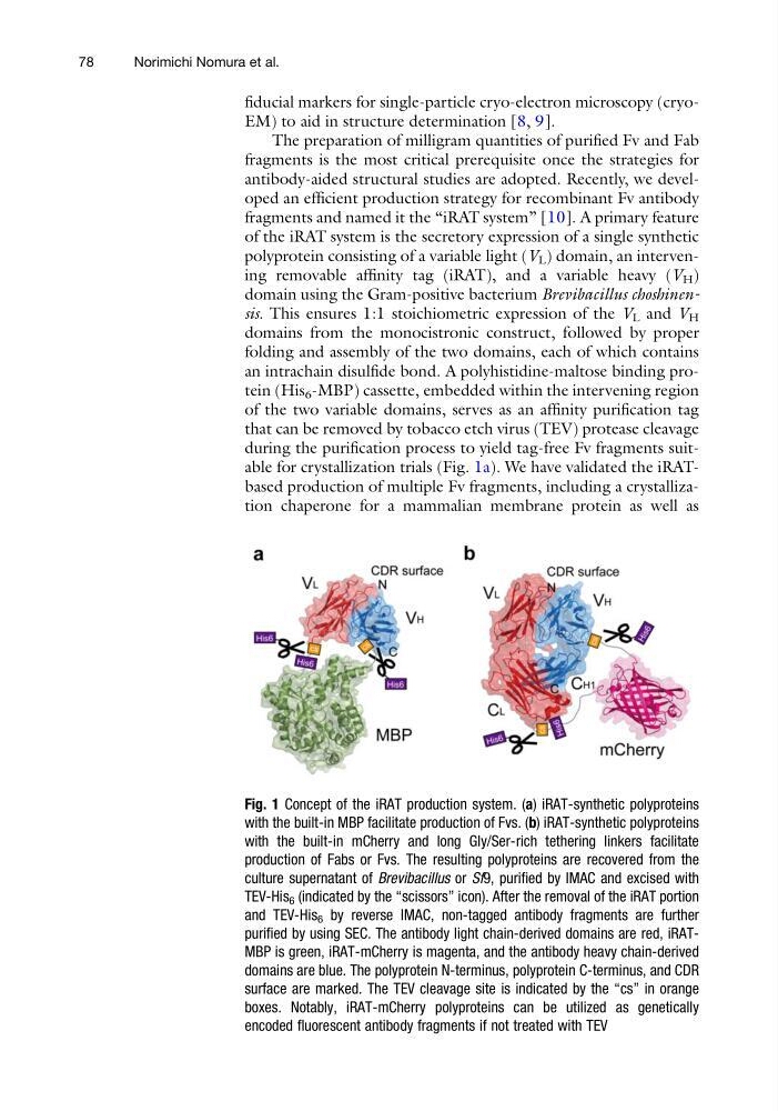
during the purification process to yield tag-free Fv fragments suitable for crystallization trials (Fig. 1a). We have validated the iRATbased production of multiple Fv fragments, including a crystallization chaperone for a mammalian membrane protein as well as
Fig. 1 Concept of the iRAT production system. (a) iRAT-synthetic polyproteins
with the built-in MBP facilitate production of Fvs. (b) iRAT-synthetic polyproteins
with the built-in mCherry and long Gly/Ser-rich tethering linkers facilitate
production of Fabs or Fvs. The resulting polyproteins are recovered from the
culture supernatant of Brevibacillus or Sf9, purified by IMAC and excised with
TEV-His 6 (indicated by the “scissors” icon). After the removal of the iRAT portion
and TEV-His 6 by reverse IMAC, non-tagged antibody fragments are further
purified by using SEC. The antibody light chain-derived domains are red, iRATMBP is green, iRAT-mCherry is magenta, and the antibody heavy chain-derived
domains are blue. The polyprotein N-terminus, polyprotein C-terminus, and CDR
surface are marked. The TEV cleavage site is indicated by the “cs” in orange
boxes. Notably, iRAT-mCherry polyproteins can be utilized as genetically
encoded fluorescent antibody fragments if not treated with TEV
78
Norimichi Nomura et al.
The preparation of milligram quantities of purified Fv and Fab
fragments is the most critical prerequisite once the strategies for
antibody-aided structural studies are adopted. Recently, we developed an efficient production strategy for recombinant Fv antibody
fragments and named it the “iRAT system” [10]. A primary feature
of the iRAT system is the secretory expression of a single synthetic
polyprotein consisting of a variable light (V L ) domain, an intervening removable affinity tag (iRAT), and a variable heavy (V H )
domain using the Gram-positive bacterium Brevibacillus choshinensis. This ensures 1:1 stoichiometric expression of the V L and V H
domains from the monocistronic construct, followed by proper
folding and assembly of the two domains, each of which contains
an intrachain disulfide bond. A polyhistidine-maltose binding protein (His 6 -MBP) cassette, embedded within the intervening region
of the two variable domains, serves as an affinity purification tag
that can be removed by tobacco etch virus (TEV) protease cleavage
during the purification process to yield tag-free Fv fragments suitable for crystallization trials (Fig. 1a). We have validated the iRATbased production of multiple Fv fragments, including a crystallization chaperone for a mammalian membrane protein as well as
Fig. 1 Concept of the iRAT production system. (a) iRAT-synthetic polyproteins
with the built-in MBP facilitate production of Fvs. (b) iRAT-synthetic polyproteins
with the built-in mCherry and long Gly/Ser-rich tethering linkers facilitate
production of Fabs or Fvs. The resulting polyproteins are recovered from the
culture supernatant of Brevibacillus or Sf9, purified by IMAC and excised with
TEV-His 6 (indicated by the “scissors” icon). After the removal of the iRAT portion
and TEV-His 6 by reverse IMAC, non-tagged antibody fragments are further
purified by using SEC. The antibody light chain-derived domains are red, iRATMBP is green, iRAT-mCherry is magenta, and the antibody heavy chain-derived
domains are blue. The polyprotein N-terminus, polyprotein C-terminus, and CDR
surface are marked. The TEV cleavage site is indicated by the “cs” in orange
boxes. Notably, iRAT-mCherry polyproteins can be utilized as genetically
encoded fluorescent antibody fragments if not treated with TEV
78
Norimichi Nomura et al.