

1. [HisTrap Excel column chromatography] The His-tagged Fab
antibodies and proKLK7 were isolated using the A ¨ KTA start
system from (GE Healthcare) using HisTrap Excel columns for
direct loading of cell culture supernatants. The pre-filtered
(0.45 μm) supernatants were loaded onto a 1 mL HisTrap
Excel column equilibrated with buffer A (20 mM sodium
phosphate buffer, pH 7.4, 500 mM NaCl). Washing was
done using buffer A containing 10–30 mM imidazole. The
protein was eluted using buffer A containing 250 mM imidazole. Eluted fractions were analyzed by SDS-PAGE electrophoresis. Protein-containing fractions were pooled and dialyzed
against PBS using Spectra/Por dialysis tubing (Spectrum,
MWCO 6-8000) to remove imidazole from the sample. The
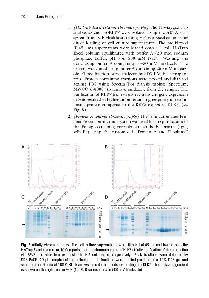
purification of KLK7 from virus-free transient gene expression
in Hi5 resulted in higher amounts and higher purity of recombinant protein compared to the BEVS expressed KLK7. (see
Fig. 5).
2. [Protein A column chromatography] The semi-automated Profinia Protein purification system was used for the purification of
the Fc-tag containing recombinant antibody formats (IgG,
scFv-Fc) using the customized “Protein A and Desalting”
Fig. 5 Affinity chromatography. The cell culture supernatants were filtrated (0.45 m) and loaded onto the
HisTrap Excel column. (a, b) Comparison of the chromatograms of KLK7 affinity purification of the production
via BEVS and virus-free expression in Hi5 cells (c, d, respectively). Peak fractions were detected by
SDS-PAGE. 20 μL samples of the collected 1 mL fractions were applied per lane of a 12% SDS-gel and
separated for 50 min at 160 V. Black arrows indicate the bands resembling pro-KLK7. The imidazole gradient
is shown on the right axis in % B (100% B corresponds to 500 mM Imidazole)
70
Jens Ko ¨ nig et al.
antibodies and proKLK7 were isolated using the A ¨ KTA start
system from (GE Healthcare) using HisTrap Excel columns for
direct loading of cell culture supernatants. The pre-filtered
(0.45 μm) supernatants were loaded onto a 1 mL HisTrap
Excel column equilibrated with buffer A (20 mM sodium
phosphate buffer, pH 7.4, 500 mM NaCl). Washing was
done using buffer A containing 10–30 mM imidazole. The
protein was eluted using buffer A containing 250 mM imidazole. Eluted fractions were analyzed by SDS-PAGE electrophoresis. Protein-containing fractions were pooled and dialyzed
against PBS using Spectra/Por dialysis tubing (Spectrum,
MWCO 6-8000) to remove imidazole from the sample. The
purification of KLK7 from virus-free transient gene expression
in Hi5 resulted in higher amounts and higher purity of recombinant protein compared to the BEVS expressed KLK7. (see
Fig. 5).
2. [Protein A column chromatography] The semi-automated Profinia Protein purification system was used for the purification of
the Fc-tag containing recombinant antibody formats (IgG,
scFv-Fc) using the customized “Protein A and Desalting”
Fig. 5 Affinity chromatography. The cell culture supernatants were filtrated (0.45 m) and loaded onto the
HisTrap Excel column. (a, b) Comparison of the chromatograms of KLK7 affinity purification of the production
via BEVS and virus-free expression in Hi5 cells (c, d, respectively). Peak fractions were detected by
SDS-PAGE. 20 μL samples of the collected 1 mL fractions were applied per lane of a 12% SDS-gel and
separated for 50 min at 160 V. Black arrows indicate the bands resembling pro-KLK7. The imidazole gradient
is shown on the right axis in % B (100% B corresponds to 500 mM Imidazole)
70
Jens Ko ¨ nig et al.