

The transfer of the scFv gene fragment into the vector
pCSE2.6-hIgG1-Fc-XP [16, 18] can be done by simple restriction
digestion and ligation using the isolated scFv phage DNA from the
library. However, for the generation of the other formats, the VH
and VL DNA fragments have to be individually isolated by PCR
amplification with the addition of new compatible cloning sites.
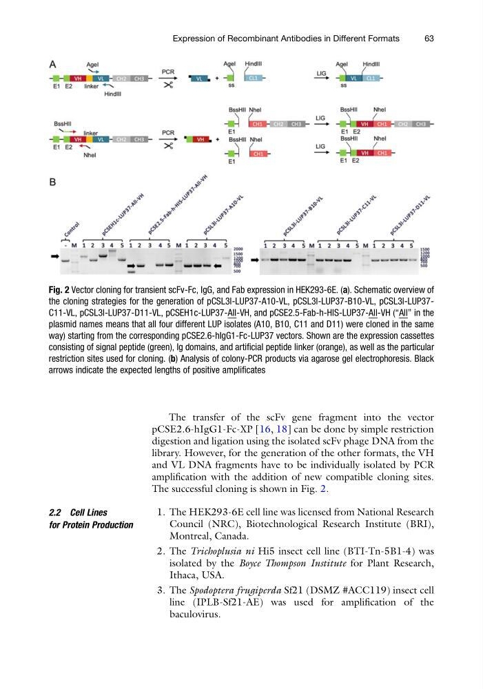
The successful cloning is shown in Fig. 2.
2.2 Cell Lines
for Protein Production
1. The HEK293-6E cell line was licensed from National Research
Council (NRC), Biotechnological Research Institute (BRI),
Montreal, Canada.
2. The Trichoplusia ni Hi5 insect cell line (BTI-Tn-5B1-4) was
isolated by the Boyce Thompson Institute for Plant Research,
Ithaca, USA.
3. The Spodoptera frugiperda Sf21 (DSMZ #ACC119) insect cell
line (IPLB-Sf21-AE) was used for amplification of the
baculovirus.
Fig. 2 Vector cloning for transient scFv-Fc, IgG, and Fab expression in HEK293-6E. (a). Schematic overview of
the cloning strategies for the generation of pCSL3l-LUP37-A10-VL, pCSL3l-LUP37-B10-VL, pCSL3l-LUP37C11-VL, pCSL3l-LUP37-D11-VL, pCSEH1c-LUP37-All-VH, and pCSE2.5-Fab-h-HIS-LUP37-All-VH (“All” in the
plasmid names means that all four different LUP isolates (A10, B10, C11 and D11) were cloned in the same
way) starting from the corresponding pCSE2.6-hIgG1-Fc-LUP37 vectors. Shown are the expression cassettes
consisting of signal peptide (green), Ig domains, and artificial peptide linker (orange), as well as the particular
restriction sites used for cloning. (b) Analysis of colony-PCR products via agarose gel electrophoresis. Black
arrows indicate the expected lengths of positive amplificates
Expression of Recombinant Antibodies in Different Formats
63
pCSE2.6-hIgG1-Fc-XP [16, 18] can be done by simple restriction
digestion and ligation using the isolated scFv phage DNA from the
library. However, for the generation of the other formats, the VH
and VL DNA fragments have to be individually isolated by PCR
amplification with the addition of new compatible cloning sites.
The successful cloning is shown in Fig. 2.
2.2 Cell Lines
for Protein Production
1. The HEK293-6E cell line was licensed from National Research
Council (NRC), Biotechnological Research Institute (BRI),
Montreal, Canada.
2. The Trichoplusia ni Hi5 insect cell line (BTI-Tn-5B1-4) was
isolated by the Boyce Thompson Institute for Plant Research,
Ithaca, USA.
3. The Spodoptera frugiperda Sf21 (DSMZ #ACC119) insect cell
line (IPLB-Sf21-AE) was used for amplification of the
baculovirus.
Fig. 2 Vector cloning for transient scFv-Fc, IgG, and Fab expression in HEK293-6E. (a). Schematic overview of
the cloning strategies for the generation of pCSL3l-LUP37-A10-VL, pCSL3l-LUP37-B10-VL, pCSL3l-LUP37C11-VL, pCSL3l-LUP37-D11-VL, pCSEH1c-LUP37-All-VH, and pCSE2.5-Fab-h-HIS-LUP37-All-VH (“All” in the
plasmid names means that all four different LUP isolates (A10, B10, C11 and D11) were cloned in the same
way) starting from the corresponding pCSE2.6-hIgG1-Fc-LUP37 vectors. Shown are the expression cassettes
consisting of signal peptide (green), Ig domains, and artificial peptide linker (orange), as well as the particular
restriction sites used for cloning. (b) Analysis of colony-PCR products via agarose gel electrophoresis. Black
arrows indicate the expected lengths of positive amplificates
Expression of Recombinant Antibodies in Different Formats
63