

1.3 Analysis
of the Binding Kinetics
of the Antibodies
and KLK7 Using
Biolayer
Interferometry (BLI)
Binding affinities of antibodies to their target protein (antigen) can
be determined by the dip and read biosensor technology of the
OctetRed System. This technology uses biolayer interferometry
(BLI) to measure protein–protein interactions [3]. The data are
generated in real time, allowing kinetic analysis as well as quantitative analysis of bound protein. Here, we describe the application to
determine the binding affinity K D of immobilized biotinylatedKLK7 to streptavidin sensors to the different antibody formats.
After initial equilibration of the biosensor, the ligand (KLK7) is
tightly bound to the tip. Binding of protein results in a change of
the thickness of the biolayer at the tip of the biosensor. The increase
in thickness of the biolayer results in a change of the interference
between two reflected beams from the internal reference layer and
the surface of the tip used for specific binding to the protein
samples. The binding of protein is detected and reported as a
change in wavelength. In this way, the association and dissociation
of the antibody to the immobilized KLK7 can be followed in time
for a range of concentrations using parallel tips. The kinetic constants can be calculated from these curves presuming a 1:1 interaction stoichiometry, which allows proper fitting of the curves.
2 Materials
In the first part, we present an overview of the vectors used, to
enable the expression of the scFv fragment derived from the phage
display analysis into different antibody formats. Furthermore, we
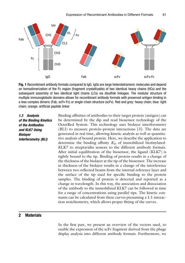
Fig. 1 Recombinant antibody formats compared to IgG. IgGs are large heterotetrameric molecules and depend
on homodimerization of the Fc region (fragment crystallizable) of two identical heavy chains (HCs) and the
subsequent assembly of two identical light chains (LCs) via disulfide linkages. The modular structure of
multiple immunoglobulin domains allows for recombinant antibody formats with preserved antigen binding in
a less complex dimeric (Fab, scFv-Fc) or single-chain structure (scFv). Red and grey: heavy chain; blue: light
chain; orange: artificial peptide linker
Expression of Recombinant Antibodies in Different Formats
61
of the Binding Kinetics
of the Antibodies
and KLK7 Using
Biolayer
Interferometry (BLI)
Binding affinities of antibodies to their target protein (antigen) can
be determined by the dip and read biosensor technology of the
OctetRed System. This technology uses biolayer interferometry
(BLI) to measure protein–protein interactions [3]. The data are
generated in real time, allowing kinetic analysis as well as quantitative analysis of bound protein. Here, we describe the application to
determine the binding affinity K D of immobilized biotinylatedKLK7 to streptavidin sensors to the different antibody formats.
After initial equilibration of the biosensor, the ligand (KLK7) is
tightly bound to the tip. Binding of protein results in a change of
the thickness of the biolayer at the tip of the biosensor. The increase
in thickness of the biolayer results in a change of the interference
between two reflected beams from the internal reference layer and
the surface of the tip used for specific binding to the protein
samples. The binding of protein is detected and reported as a
change in wavelength. In this way, the association and dissociation
of the antibody to the immobilized KLK7 can be followed in time
for a range of concentrations using parallel tips. The kinetic constants can be calculated from these curves presuming a 1:1 interaction stoichiometry, which allows proper fitting of the curves.
2 Materials
In the first part, we present an overview of the vectors used, to
enable the expression of the scFv fragment derived from the phage
display analysis into different antibody formats. Furthermore, we
Fig. 1 Recombinant antibody formats compared to IgG. IgGs are large heterotetrameric molecules and depend
on homodimerization of the Fc region (fragment crystallizable) of two identical heavy chains (HCs) and the
subsequent assembly of two identical light chains (LCs) via disulfide linkages. The modular structure of
multiple immunoglobulin domains allows for recombinant antibody formats with preserved antigen binding in
a less complex dimeric (Fab, scFv-Fc) or single-chain structure (scFv). Red and grey: heavy chain; blue: light
chain; orange: artificial peptide linker
Expression of Recombinant Antibodies in Different Formats
61