

1. Three days after transfection, wash cells with PBS, extract
genomic DNA (typically from 1 Â 10
5 cells) using QuickExtract DNA extraction kit or similar according to the manufacturer’s recommendations. Dilute genomic DNA at a final
concentration around 50 ng/μL.
2. Design a pair of PCR primers (XPB-FW and XPB-RV) which
hybridize on either side of the target site to amplify a ~500 bp
stretch of DNA. The sgRNA cut site should ideally be located
~200 bp downstream from one of the primers for convenient
analysis by DNA sequencing.
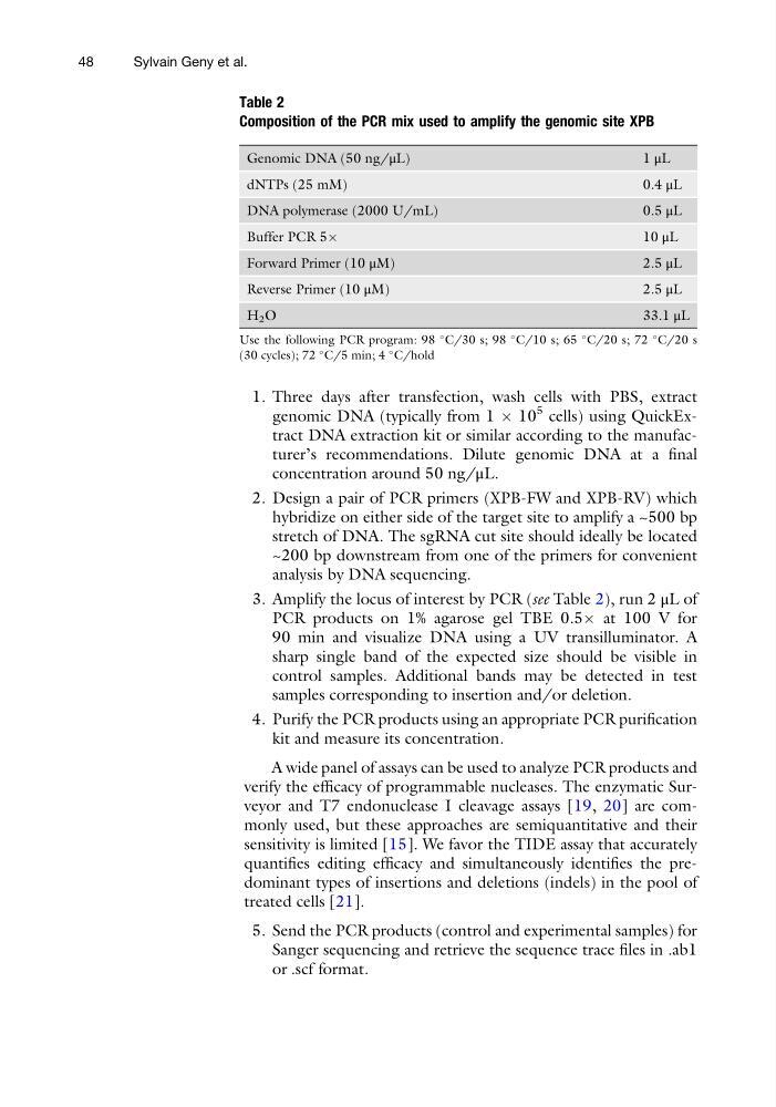
3. Amplify the locus of interest by PCR (see Table 2), run 2 μL of
PCR products on 1% agarose gel TBE 0.5Â at 100 V for
90 min and visualize DNA using a UV transilluminator. A
sharp single band of the expected size should be visible in
control samples. Additional bands may be detected in test
samples corresponding to insertion and/or deletion.
4. Purify the PCR products using an appropriate PCR purification
kit and measure its concentration.
A wide panel of assays can be used to analyze PCR products and
verify the efficacy of programmable nucleases. The enzymatic Surveyor and T7 endonuclease I cleavage assays [19, 20] are commonly used, but these approaches are semiquantitative and their
sensitivity is limited [15]. We favor the TIDE assay that accurately
quantifies editing efficacy and simultaneously identifies the predominant types of insertions and deletions (indels) in the pool of
treated cells [21].
5. Send the PCR products (control and experimental samples) for
Sanger sequencing and retrieve the sequence trace files in .ab1
or .scf format.
Table 2
Composition of the PCR mix used to amplify the genomic site XPB
Genomic DNA (50 ng/μL)
1 μL
dNTPs (25 mM)
0.4 μL
DNA polymerase (2000 U/mL)
0.5 μL
Buffer PCR 5Â
10 μL
Forward Primer (10 μM)
2.5 μL
Reverse Primer (10 μM)
2.5 μL
H 2 O
33.1 μL
Use the following PCR program: 98
C/30 s; 98
C/10 s; 65
C/20 s; 72
C/20 s
(30 cycles); 72
C/5 min; 4
C/hold
48
Sylvain Geny et al.
genomic DNA (typically from 1 Â 10
5 cells) using QuickExtract DNA extraction kit or similar according to the manufacturer’s recommendations. Dilute genomic DNA at a final
concentration around 50 ng/μL.
2. Design a pair of PCR primers (XPB-FW and XPB-RV) which
hybridize on either side of the target site to amplify a ~500 bp
stretch of DNA. The sgRNA cut site should ideally be located
~200 bp downstream from one of the primers for convenient
analysis by DNA sequencing.
3. Amplify the locus of interest by PCR (see Table 2), run 2 μL of
PCR products on 1% agarose gel TBE 0.5Â at 100 V for
90 min and visualize DNA using a UV transilluminator. A
sharp single band of the expected size should be visible in
control samples. Additional bands may be detected in test
samples corresponding to insertion and/or deletion.
4. Purify the PCR products using an appropriate PCR purification
kit and measure its concentration.
A wide panel of assays can be used to analyze PCR products and
verify the efficacy of programmable nucleases. The enzymatic Surveyor and T7 endonuclease I cleavage assays [19, 20] are commonly used, but these approaches are semiquantitative and their
sensitivity is limited [15]. We favor the TIDE assay that accurately
quantifies editing efficacy and simultaneously identifies the predominant types of insertions and deletions (indels) in the pool of
treated cells [21].
5. Send the PCR products (control and experimental samples) for
Sanger sequencing and retrieve the sequence trace files in .ab1
or .scf format.
Table 2
Composition of the PCR mix used to amplify the genomic site XPB
Genomic DNA (50 ng/μL)
1 μL
dNTPs (25 mM)
0.4 μL
DNA polymerase (2000 U/mL)
0.5 μL
Buffer PCR 5Â
10 μL
Forward Primer (10 μM)
2.5 μL
Reverse Primer (10 μM)
2.5 μL
H 2 O
33.1 μL
Use the following PCR program: 98
C/30 s; 98
C/10 s; 65
C/20 s; 72
C/20 s
(30 cycles); 72
C/5 min; 4
C/hold
48
Sylvain Geny et al.