

3.1.2 Ligation
1. Digested PCR product is pooled and ligated overnight at 16
C
(see Note 4).
Ligation(scale to adjust
DNA concentration <10 ng/μl)
ddH 2 O
325 μL
Ligation buffer (10Â)
5 0 μL
Digested PCR product
100 μL
T4 ligase (5 U μL
À1 )
2 5 μL
Total volume
500 μL
2. Precipitate ligation reaction with ethanol by addition of 1/10
vol. of 3 M NaOAc, pH 5.2–500 μL of ligation reaction and
2 vol. of 100% ethanol.
3. Let the solution stand on ice for 15 min.
4. Centrifuge at 19,000 Â g for 45 min at 4
C (see Note 5).
5. Remove the supernatant.
6. Add 1 mL of 70% ethanol.
7. Vortex shortly and let stand on ice for 10 min.
8. Centrifuge at 19,000 Â g for 45 min at 4
C.
9. Repeat the last two steps one more time.
10. Remove the ethanol.
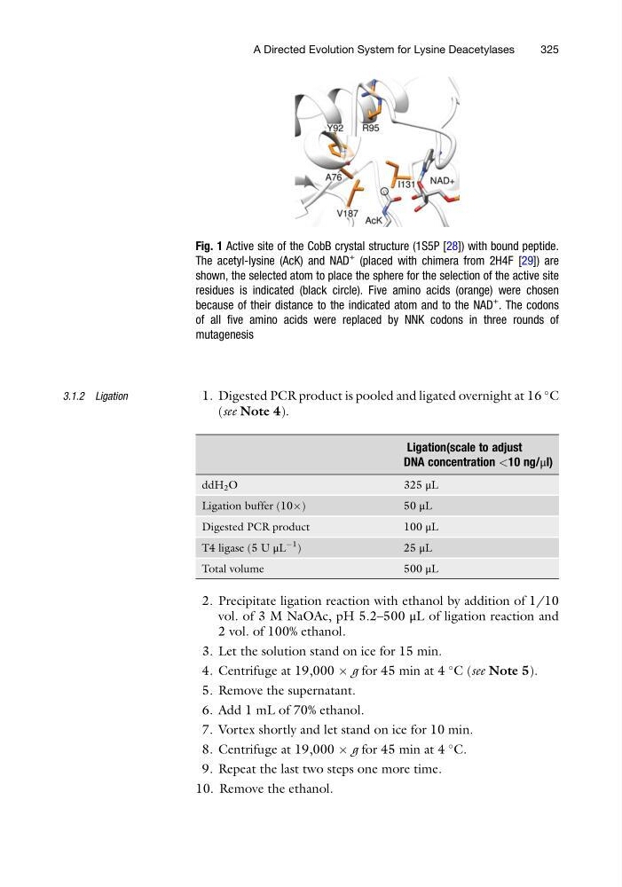
Fig. 1 Active site of the CobB crystal structure (1S5P [28]) with bound peptide.
The acetyl-lysine (AcK) and NAD
+
(placed with chimera from 2H4F [29]) are
shown, the selected atom to place the sphere for the selection of the active site
residues is indicated (black circle). Five amino acids (orange) were chosen
because of their distance to the indicated atom and to the NAD
+
. The codons
of all five amino acids were replaced by NNK codons in three rounds of
mutagenesis
A Directed Evolution System for Lysine Deacetylases
325
1. Digested PCR product is pooled and ligated overnight at 16
C
(see Note 4).
Ligation(scale to adjust
DNA concentration <10 ng/μl)
ddH 2 O
325 μL
Ligation buffer (10Â)
5 0 μL
Digested PCR product
100 μL
T4 ligase (5 U μL
À1 )
2 5 μL
Total volume
500 μL
2. Precipitate ligation reaction with ethanol by addition of 1/10
vol. of 3 M NaOAc, pH 5.2–500 μL of ligation reaction and
2 vol. of 100% ethanol.
3. Let the solution stand on ice for 15 min.
4. Centrifuge at 19,000 Â g for 45 min at 4
C (see Note 5).
5. Remove the supernatant.
6. Add 1 mL of 70% ethanol.
7. Vortex shortly and let stand on ice for 10 min.
8. Centrifuge at 19,000 Â g for 45 min at 4
C.
9. Repeat the last two steps one more time.
10. Remove the ethanol.
Fig. 1 Active site of the CobB crystal structure (1S5P [28]) with bound peptide.
The acetyl-lysine (AcK) and NAD
+
(placed with chimera from 2H4F [29]) are
shown, the selected atom to place the sphere for the selection of the active site
residues is indicated (black circle). Five amino acids (orange) were chosen
because of their distance to the indicated atom and to the NAD
+
. The codons
of all five amino acids were replaced by NNK codons in three rounds of
mutagenesis
A Directed Evolution System for Lysine Deacetylases
325