

the fusion to CBirA*) diluted at their optimal concentrations
(see Note 19) in PBS containing 2% bovine serum albumin
(BSA) and 0.1% Tween-20 for 1 h at RT on a roller-shaker (see
Note 20).
16. Wash the membrane three times with PBS containing 0.1%
Tween-20 for 10 min at RT.
17. Incubate the membrane in a 50-mL falcon tube with 5 mL of
conjugated secondary antibodies directed against the anti-Myc
and -FLAG primary antibodies diluted at their optimal concentrations (see Note 21) in PBS containing 2% bovine serum
albumin (BSA) and 0.1% Tween-20 for 30 min at RT on a
roller-shaker.
18. Wash the membrane three times with PBS containing 0.1%
Tween-20 for 10 min at RT (see Note 18) and then proceed
with detection of chemoluminescence or fluorescence signals.
3.3 Large-Scale
Proximity-Dependent
Biotinylation
for Proteomics Studies
1. For each condition to be tested, seed three to four 10-cm cell
culture dishes with the cells to transfect, so that they reach
approximately 70% confluence on the following day. Incubate
overnight at 37
C, 5% CO 2 . For HeLa cells, typically 8 Â 10
5
cells are seeded per dish in a volume of 10 mL (see Note 14).
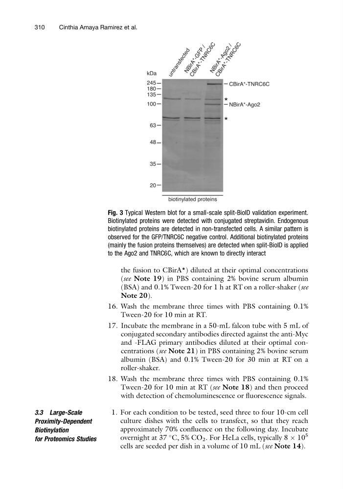
Fig. 3 Typical Western blot for a small-scale split-BioID validation experiment.
Biotinylated proteins were detected with conjugated streptavidin. Endogenous
biotinylated proteins are detected in non-transfected cells. A similar pattern is
observed for the GFP/TNRC6C negative control. Additional biotinylated proteins
(mainly the fusion proteins themselves) are detected when split-BioID is applied
to the Ago2 and TNRC6C, which are known to directly interact
310
Cinthia Amaya Ramirez et al.
(see Note 19) in PBS containing 2% bovine serum albumin
(BSA) and 0.1% Tween-20 for 1 h at RT on a roller-shaker (see
Note 20).
16. Wash the membrane three times with PBS containing 0.1%
Tween-20 for 10 min at RT.
17. Incubate the membrane in a 50-mL falcon tube with 5 mL of
conjugated secondary antibodies directed against the anti-Myc
and -FLAG primary antibodies diluted at their optimal concentrations (see Note 21) in PBS containing 2% bovine serum
albumin (BSA) and 0.1% Tween-20 for 30 min at RT on a
roller-shaker.
18. Wash the membrane three times with PBS containing 0.1%
Tween-20 for 10 min at RT (see Note 18) and then proceed
with detection of chemoluminescence or fluorescence signals.
3.3 Large-Scale
Proximity-Dependent
Biotinylation
for Proteomics Studies
1. For each condition to be tested, seed three to four 10-cm cell
culture dishes with the cells to transfect, so that they reach
approximately 70% confluence on the following day. Incubate
overnight at 37
C, 5% CO 2 . For HeLa cells, typically 8 Â 10
5
cells are seeded per dish in a volume of 10 mL (see Note 14).
Fig. 3 Typical Western blot for a small-scale split-BioID validation experiment.
Biotinylated proteins were detected with conjugated streptavidin. Endogenous
biotinylated proteins are detected in non-transfected cells. A similar pattern is
observed for the GFP/TNRC6C negative control. Additional biotinylated proteins
(mainly the fusion proteins themselves) are detected when split-BioID is applied
to the Ago2 and TNRC6C, which are known to directly interact
310
Cinthia Amaya Ramirez et al.