

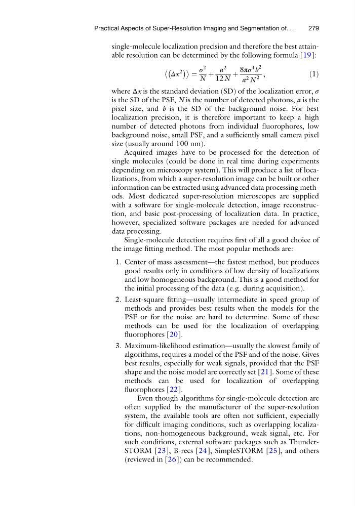
single-molecule localization precision and therefore the best attainable resolution can be determined by the following formula [19]:
Δx
2
À
Á
¼
σ
2
N
þ
a
2
12N
þ
8πσ
4
b
2
a 2 N
2
,
ð1Þ
where Δx is the standard deviation (SD) of the localization error, σ
is the SD of the PSF, N is the number of detected photons, a is the
pixel size, and b is the SD of the background noise. For best
localization precision, it is therefore important to keep a high
number of detected photons from individual fluorophores, low
background noise, small PSF, and a sufficiently small camera pixel
size (usually around 100 nm).
Acquired images have to be processed for the detection of
single molecules (could be done in real time during experiments
depending on microscopy system). This will produce a list of localizations, from which a super-resolution image can be built or other
information can be extracted using advanced data processing methods. Most dedicated super-resolution microscopes are supplied
with a software for single-molecule detection, image reconstruction, and basic post-processing of localization data. In practice,
however, specialized software packages are needed for advanced
data processing.
Single-molecule detection requires first of all a good choice of
the image fitting method. The most popular methods are:
1. Center of mass assessment—the fastest method, but produces
good results only in conditions of low density of localizations
and low homogeneous background. This is a good method for
the initial processing of the data (e.g. during acquisition).
2. Least-square fitting—usually intermediate in speed group of
methods and provides best results when the models for the
PSF or for the noise are hard to determine. Some of these
methods can be used for the localization of overlapping
fluorophores [20].
3. Maximum-likelihood estimation—usually the slowest family of
algorithms, requires a model of the PSF and of the noise. Gives
best results, especially for weak signals, provided that the PSF
shape and the noise model are correctly set [21]. Some of these
methods can be used for localization of overlapping
fluorophores [22].
Even though algorithms for single-molecule detection are
often supplied by the manufacturer of the super-resolution
system, the available tools are often not sufficient, especially
for difficult imaging conditions, such as overlapping localizations, non-homogeneous background, weak signal, etc. For
such conditions, external software packages such as ThunderSTORM [23], B-recs [24], SimpleSTORM [25], and others
(reviewed in [26]) can be recommended.
Practical Aspects of Super-Resolution Imaging and Segmentation of. . .
279
Δx
2
À
Á
¼
σ
2
N
þ
a
2
12N
þ
8πσ
4
b
2
a 2 N
2
,
ð1Þ
where Δx is the standard deviation (SD) of the localization error, σ
is the SD of the PSF, N is the number of detected photons, a is the
pixel size, and b is the SD of the background noise. For best
localization precision, it is therefore important to keep a high
number of detected photons from individual fluorophores, low
background noise, small PSF, and a sufficiently small camera pixel
size (usually around 100 nm).
Acquired images have to be processed for the detection of
single molecules (could be done in real time during experiments
depending on microscopy system). This will produce a list of localizations, from which a super-resolution image can be built or other
information can be extracted using advanced data processing methods. Most dedicated super-resolution microscopes are supplied
with a software for single-molecule detection, image reconstruction, and basic post-processing of localization data. In practice,
however, specialized software packages are needed for advanced
data processing.
Single-molecule detection requires first of all a good choice of
the image fitting method. The most popular methods are:
1. Center of mass assessment—the fastest method, but produces
good results only in conditions of low density of localizations
and low homogeneous background. This is a good method for
the initial processing of the data (e.g. during acquisition).
2. Least-square fitting—usually intermediate in speed group of
methods and provides best results when the models for the
PSF or for the noise are hard to determine. Some of these
methods can be used for the localization of overlapping
fluorophores [20].
3. Maximum-likelihood estimation—usually the slowest family of
algorithms, requires a model of the PSF and of the noise. Gives
best results, especially for weak signals, provided that the PSF
shape and the noise model are correctly set [21]. Some of these
methods can be used for localization of overlapping
fluorophores [22].
Even though algorithms for single-molecule detection are
often supplied by the manufacturer of the super-resolution
system, the available tools are often not sufficient, especially
for difficult imaging conditions, such as overlapping localizations, non-homogeneous background, weak signal, etc. For
such conditions, external software packages such as ThunderSTORM [23], B-recs [24], SimpleSTORM [25], and others
(reviewed in [26]) can be recommended.
Practical Aspects of Super-Resolution Imaging and Segmentation of. . .
279