

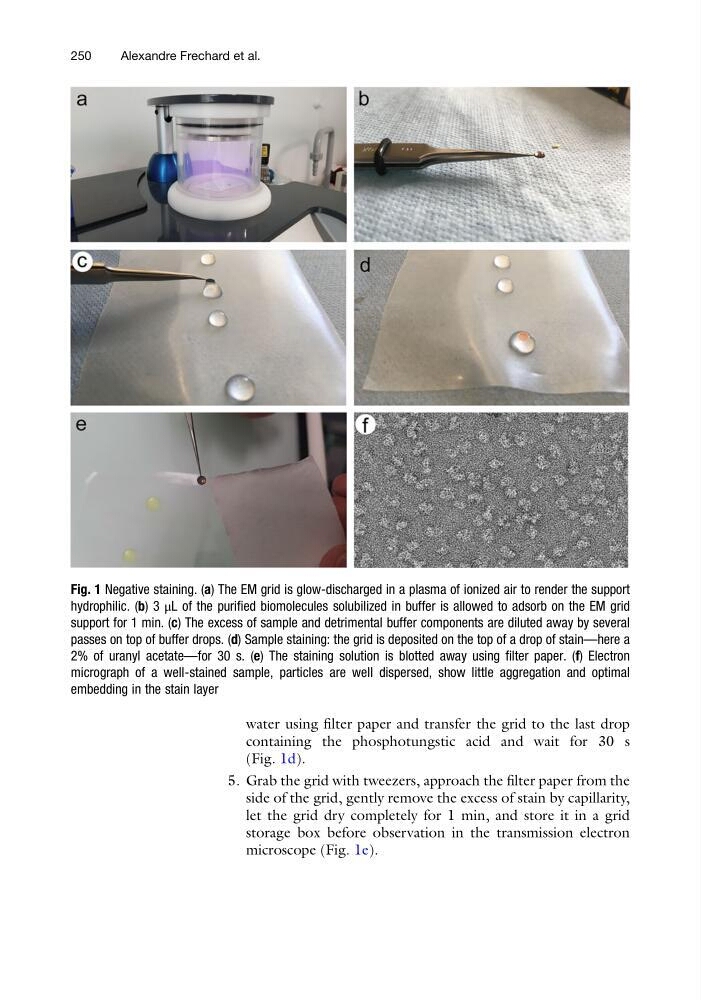
water using filter paper and transfer the grid to the last drop
containing the phosphotungstic acid and wait for 30 s
(Fig. 1d).
5. Grab the grid with tweezers, approach the filter paper from the
side of the grid, gently remove the excess of stain by capillarity,
let the grid dry completely for 1 min, and store it in a grid
storage box before observation in the transmission electron
microscope (Fig. 1e).
Fig. 1 Negative staining. (a) The EM grid is glow-discharged in a plasma of ionized air to render the support
hydrophilic. (b) 3 μL of the purified biomolecules solubilized in buffer is allowed to adsorb on the EM grid
support for 1 min. (c) The excess of sample and detrimental buffer components are diluted away by several
passes on top of buffer drops. (d) Sample staining: the grid is deposited on the top of a drop of stain—here a
2% of uranyl acetate—for 30 s. (e) The staining solution is blotted away using filter paper. (f) Electron
micrograph of a well-stained sample, particles are well dispersed, show little aggregation and optimal
embedding in the stain layer
250
Alexandre Frechard et al.
containing the phosphotungstic acid and wait for 30 s
(Fig. 1d).
5. Grab the grid with tweezers, approach the filter paper from the
side of the grid, gently remove the excess of stain by capillarity,
let the grid dry completely for 1 min, and store it in a grid
storage box before observation in the transmission electron
microscope (Fig. 1e).
Fig. 1 Negative staining. (a) The EM grid is glow-discharged in a plasma of ionized air to render the support
hydrophilic. (b) 3 μL of the purified biomolecules solubilized in buffer is allowed to adsorb on the EM grid
support for 1 min. (c) The excess of sample and detrimental buffer components are diluted away by several
passes on top of buffer drops. (d) Sample staining: the grid is deposited on the top of a drop of stain—here a
2% of uranyl acetate—for 30 s. (e) The staining solution is blotted away using filter paper. (f) Electron
micrograph of a well-stained sample, particles are well dispersed, show little aggregation and optimal
embedding in the stain layer
250
Alexandre Frechard et al.