

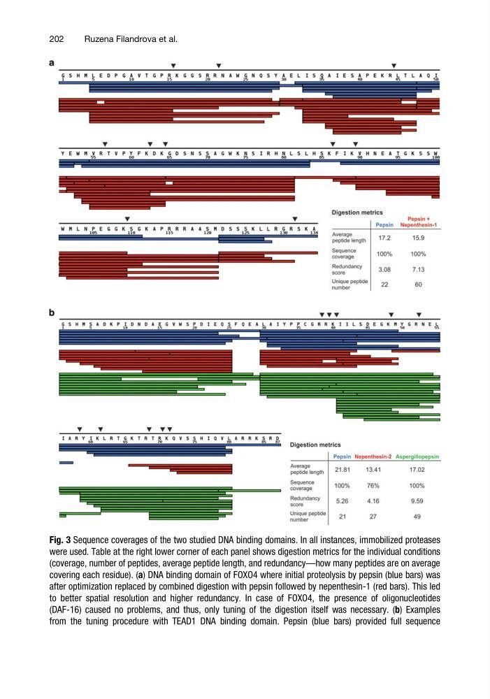
Fig. 3 Sequence coverages of the two studied DNA binding domains. In all instances, immobilized proteases
were used. Table at the right lower corner of each panel shows digestion metrics for the individual conditions
(coverage, number of peptides, average peptide length, and redundancy—how many peptides are on average
covering each residue). (a) DNA binding domain of FOXO4 where initial proteolysis by pepsin (blue bars) was
after optimization replaced by combined digestion with pepsin followed by nepenthesin-1 (red bars). This led
to better spatial resolution and higher redundancy. In case of FOXO4, the presence of oligonucleotides
(DAF-16) caused no problems, and thus, only tuning of the digestion itself was necessary. (b) Examples
from the tuning procedure with TEAD1 DNA binding domain. Pepsin (blue bars) provided full sequence
202
Ruzena Filandrova et al.
were used. Table at the right lower corner of each panel shows digestion metrics for the individual conditions
(coverage, number of peptides, average peptide length, and redundancy—how many peptides are on average
covering each residue). (a) DNA binding domain of FOXO4 where initial proteolysis by pepsin (blue bars) was
after optimization replaced by combined digestion with pepsin followed by nepenthesin-1 (red bars). This led
to better spatial resolution and higher redundancy. In case of FOXO4, the presence of oligonucleotides
(DAF-16) caused no problems, and thus, only tuning of the digestion itself was necessary. (b) Examples
from the tuning procedure with TEAD1 DNA binding domain. Pepsin (blue bars) provided full sequence
202
Ruzena Filandrova et al.