

proteolytic activity [17]. In addition, guanidine is difficult to
remove completely during the subsequent desalting step, and
thus, 4 M urea (final concentration upon quenching) may always
be suggested as the first choice.
When analyzing samples containing nucleic acids, specific problems may additionally arise upon the quenching of H/D
exchange, as mentioned above. As the pH drops, nucleic acids
tend to precipitate, and it is very likely that the protein of interest
will coprecipitate as well. Therefore, several strategies described in
the Introduction were developed to remove the nucleic acids after
quenching or to reduce their detrimental effect on the HDX-MS
workflow.
1. Prepare the duplex DNA according to the steps 2 and 3 of the
previous chapter (Subheading 3.1) and mix it with the protein
in equimolar ratio and the concentration that will later be used
during D 2 O labeling. Incubate the mixture for 10–20 min to
ensure binding equilibrium. Also, prepare a sample without
DNA for the comparison of how DNA affects the digestion
pattern. In this step, use normal (H 2 O, not D 2 O based)
buffers.
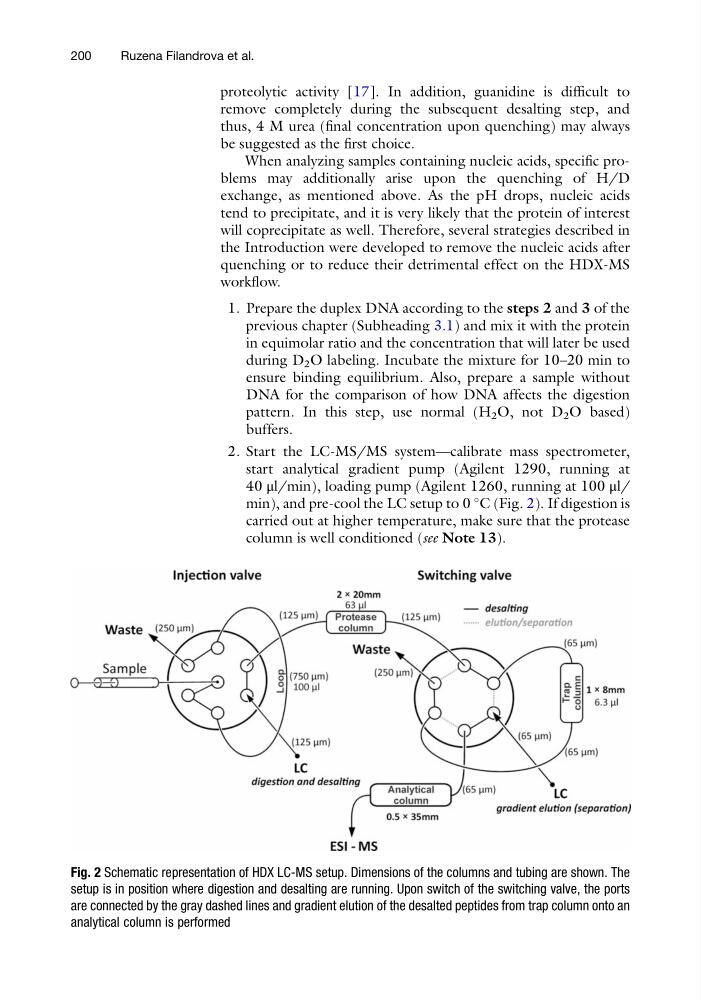
2. Start the LC-MS/MS system—calibrate mass spectrometer,
start analytical gradient pump (Agilent 1290, running at
40 μl/min), loading pump (Agilent 1260, running at 100 μl/
min), and pre-cool the LC setup to 0
C (Fig. 2). If digestion is
carried out at higher temperature, make sure that the protease
column is well conditioned (see Note 13).
Fig. 2 Schematic representation of HDX LC-MS setup. Dimensions of the columns and tubing are shown. The
setup is in position where digestion and desalting are running. Upon switch of the switching valve, the ports
are connected by the gray dashed lines and gradient elution of the desalted peptides from trap column onto an
analytical column is performed
200
Ruzena Filandrova et al.
remove completely during the subsequent desalting step, and
thus, 4 M urea (final concentration upon quenching) may always
be suggested as the first choice.
When analyzing samples containing nucleic acids, specific problems may additionally arise upon the quenching of H/D
exchange, as mentioned above. As the pH drops, nucleic acids
tend to precipitate, and it is very likely that the protein of interest
will coprecipitate as well. Therefore, several strategies described in
the Introduction were developed to remove the nucleic acids after
quenching or to reduce their detrimental effect on the HDX-MS
workflow.
1. Prepare the duplex DNA according to the steps 2 and 3 of the
previous chapter (Subheading 3.1) and mix it with the protein
in equimolar ratio and the concentration that will later be used
during D 2 O labeling. Incubate the mixture for 10–20 min to
ensure binding equilibrium. Also, prepare a sample without
DNA for the comparison of how DNA affects the digestion
pattern. In this step, use normal (H 2 O, not D 2 O based)
buffers.
2. Start the LC-MS/MS system—calibrate mass spectrometer,
start analytical gradient pump (Agilent 1290, running at
40 μl/min), loading pump (Agilent 1260, running at 100 μl/
min), and pre-cool the LC setup to 0
C (Fig. 2). If digestion is
carried out at higher temperature, make sure that the protease
column is well conditioned (see Note 13).
Fig. 2 Schematic representation of HDX LC-MS setup. Dimensions of the columns and tubing are shown. The
setup is in position where digestion and desalting are running. Upon switch of the switching valve, the ports
are connected by the gray dashed lines and gradient elution of the desalted peptides from trap column onto an
analytical column is performed
200
Ruzena Filandrova et al.