

3. Typically, it takes 6–7 h to reach the desired OD 600nm .
4. We usually screen 12–24 neomycin-resistant colonies. The
positive rate is approximately 65–70%.
5. The built-in mCherry within the iRAT segment can be replaced
with another fluorescent protein such as EGFP or mBanana,
depending on the experimental design.
6. Bypassing the papain digestion process is one technical advantage of the iRAT-mediated Fab production. In conventional
preparation for Fabs from whole IgG, papain digestion often
results in a combination of under- and over-digestion and
requires specific optimization depending on different IgG
subclasses.
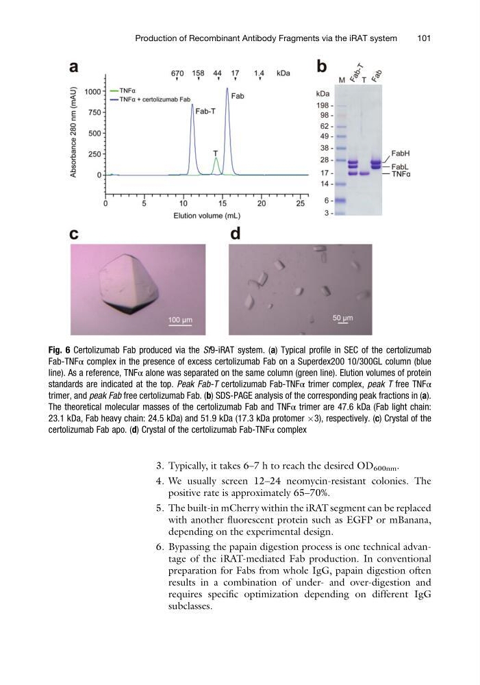
Fig. 6 Certolizumab Fab produced via the Sf9-iRAT system. (a) Typical profile in SEC of the certolizumab
Fab-TNFα complex in the presence of excess certolizumab Fab on a Superdex200 10/300GL column (blue
line). As a reference, TNFα alone was separated on the same column (green line). Elution volumes of protein
standards are indicated at the top. Peak Fab-T certolizumab Fab-TNFα trimer complex, peak T free TNFα
trimer, and peak Fab free certolizumab Fab. (b) SDS-PAGE analysis of the corresponding peak fractions in (a).
The theoretical molecular masses of the certolizumab Fab and TNFα trimer are 47.6 kDa (Fab light chain:
23.1 kDa, Fab heavy chain: 24.5 kDa) and 51.9 kDa (17.3 kDa protomer Â3), respectively. (c) Crystal of the
certolizumab Fab apo. (d) Crystal of the certolizumab Fab-TNFα complex
Production of Recombinant Antibody Fragments via the iRAT system
101
4. We usually screen 12–24 neomycin-resistant colonies. The
positive rate is approximately 65–70%.
5. The built-in mCherry within the iRAT segment can be replaced
with another fluorescent protein such as EGFP or mBanana,
depending on the experimental design.
6. Bypassing the papain digestion process is one technical advantage of the iRAT-mediated Fab production. In conventional
preparation for Fabs from whole IgG, papain digestion often
results in a combination of under- and over-digestion and
requires specific optimization depending on different IgG
subclasses.
Fig. 6 Certolizumab Fab produced via the Sf9-iRAT system. (a) Typical profile in SEC of the certolizumab
Fab-TNFα complex in the presence of excess certolizumab Fab on a Superdex200 10/300GL column (blue
line). As a reference, TNFα alone was separated on the same column (green line). Elution volumes of protein
standards are indicated at the top. Peak Fab-T certolizumab Fab-TNFα trimer complex, peak T free TNFα
trimer, and peak Fab free certolizumab Fab. (b) SDS-PAGE analysis of the corresponding peak fractions in (a).
The theoretical molecular masses of the certolizumab Fab and TNFα trimer are 47.6 kDa (Fab light chain:
23.1 kDa, Fab heavy chain: 24.5 kDa) and 51.9 kDa (17.3 kDa protomer Â3), respectively. (c) Crystal of the
certolizumab Fab apo. (d) Crystal of the certolizumab Fab-TNFα complex
Production of Recombinant Antibody Fragments via the iRAT system
101