

27. Re-measure the protein concentration of each fraction using a
NanoDrop spectrophotometer.
28. Prepare 100 μL aliquots and flash-freeze with liquid nitrogen.
29. Store at À80
C.
3.2 Extended iRAT
System
for the Production
of Recombinant Fab
Fragments and Their
Fluorescent
Derivatives
Fab fragments (with a molecular weight of ~50 kDa) are heterodimeric molecules consisting of two chains, each of which has two
domains: the light chain variable domain (V L ) linked to the light
chain constant domain (C L ) and the heavy chain variable domain
(V H ) linked to the heavy chain constant domain 1 (C H 1). Fab
fragments contain five disulfide bonds (except IgA and IgE which
have six disulfides) and need to be targeted to the Gram-negative
bacterial periplasm, the Gram-positive bacterial extracellular space,
or the eukaryotic endoplasmic reticulum. The linear distance
between the C-terminus of the Fab light chain (Fab L ) and
N-terminus of the Fab heavy chain (Fab H ) for most Fab fragments
is approximately 65 A ˚ . Appropriate long flexible linkers are needed
for successful “molecular surgery” to anastomose the two termini
without violating the overall fold and biological function of the Fab
fragments. According to Koerber et al. a 70-residue Gly/Ser-rich
linker (sc70) is available to express functional single-chain Fab
[13]. Here we provide an example of which the mCherry red
fluorescent protein, instead of MBP, is installed into an intermediate point of the sc70 linker to create a new iRAT segment. This
construct yields a polyprotein that works as a genetically encoded
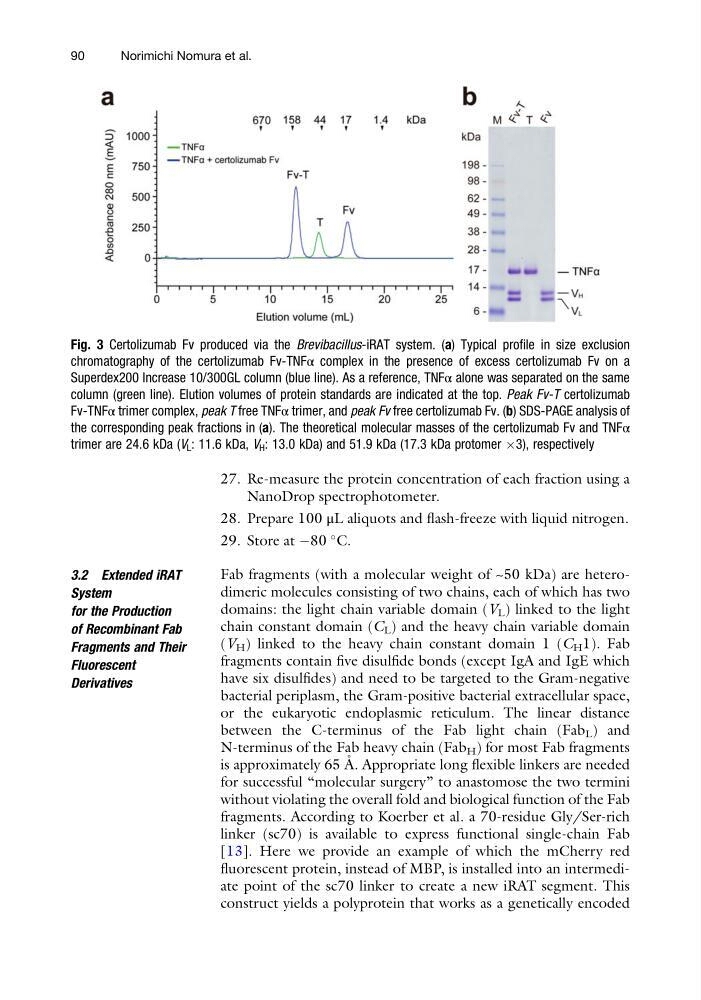
Fig. 3 Certolizumab Fv produced via the Brevibacillus-iRAT system. (a) Typical profile in size exclusion
chromatography of the certolizumab Fv-TNFα complex in the presence of excess certolizumab Fv on a
Superdex200 Increase 10/300GL column (blue line). As a reference, TNFα alone was separated on the same
column (green line). Elution volumes of protein standards are indicated at the top. Peak Fv-T certolizumab
Fv-TNFα trimer complex, peak T free TNFα trimer, and peak Fv free certolizumab Fv. (b) SDS-PAGE analysis of
the corresponding peak fractions in (a). The theoretical molecular masses of the certolizumab Fv and TNFα
trimer are 24.6 kDa (V L : 11.6 kDa, V H : 13.0 kDa) and 51.9 kDa (17.3 kDa protomer Â3), respectively
90
Norimichi Nomura et al.
NanoDrop spectrophotometer.
28. Prepare 100 μL aliquots and flash-freeze with liquid nitrogen.
29. Store at À80
C.
3.2 Extended iRAT
System
for the Production
of Recombinant Fab
Fragments and Their
Fluorescent
Derivatives
Fab fragments (with a molecular weight of ~50 kDa) are heterodimeric molecules consisting of two chains, each of which has two
domains: the light chain variable domain (V L ) linked to the light
chain constant domain (C L ) and the heavy chain variable domain
(V H ) linked to the heavy chain constant domain 1 (C H 1). Fab
fragments contain five disulfide bonds (except IgA and IgE which
have six disulfides) and need to be targeted to the Gram-negative
bacterial periplasm, the Gram-positive bacterial extracellular space,
or the eukaryotic endoplasmic reticulum. The linear distance
between the C-terminus of the Fab light chain (Fab L ) and
N-terminus of the Fab heavy chain (Fab H ) for most Fab fragments
is approximately 65 A ˚ . Appropriate long flexible linkers are needed
for successful “molecular surgery” to anastomose the two termini
without violating the overall fold and biological function of the Fab
fragments. According to Koerber et al. a 70-residue Gly/Ser-rich
linker (sc70) is available to express functional single-chain Fab
[13]. Here we provide an example of which the mCherry red
fluorescent protein, instead of MBP, is installed into an intermediate point of the sc70 linker to create a new iRAT segment. This
construct yields a polyprotein that works as a genetically encoded
Fig. 3 Certolizumab Fv produced via the Brevibacillus-iRAT system. (a) Typical profile in size exclusion
chromatography of the certolizumab Fv-TNFα complex in the presence of excess certolizumab Fv on a
Superdex200 Increase 10/300GL column (blue line). As a reference, TNFα alone was separated on the same
column (green line). Elution volumes of protein standards are indicated at the top. Peak Fv-T certolizumab
Fv-TNFα trimer complex, peak T free TNFα trimer, and peak Fv free certolizumab Fv. (b) SDS-PAGE analysis of
the corresponding peak fractions in (a). The theoretical molecular masses of the certolizumab Fv and TNFα
trimer are 24.6 kDa (V L : 11.6 kDa, V H : 13.0 kDa) and 51.9 kDa (17.3 kDa protomer Â3), respectively
90
Norimichi Nomura et al.