

3 Pulse Oximetry for the Measurement of Oxygen Saturation …
57
Fig. 3.1 Sensor for
obtaining a PPG signal
utilizing the transmitted light
through finger
IIN
I o
suitable photo detector placed on the side opposite to that of the source as indicated
in Fig. 3.1.
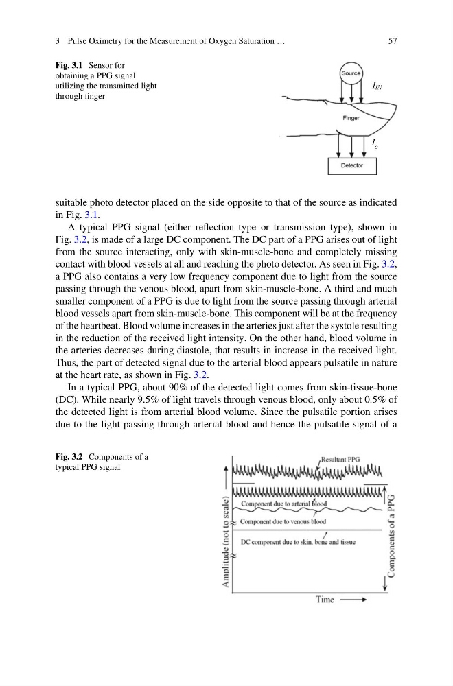
A typical PPG signal (either reflection type or transmission type), shown in
Fig. 3.2, is made of a large DC component. The DC part of a PPG arises out of light
from the source interacting, only with skin-muscle-bone and completely missing
contact with blood vessels at all and reaching the photo detector. As seen in Fig. 3.2,
a PPG also contains a very low frequency component due to light from the source
passing through the venous blood, apart from skin-muscle-bone. A third and much
smaller component of a PPG is due to light from the source passing through arterial
blood vessels apart from skin-muscle-bone. This component will be at the frequency
of the heartbeat. Blood volume increases in the arteries just after the systole resulting
in the reduction of the received light intensity. On the other hand, blood volume in
the arteries decreases during diastole, that results in increase in the received light.
Thus, the part of detected signal due to the arterial blood appears pulsatile in nature
at the heart rate, as shown in Fig. 3.2.
In a typical PPG, about 90% of the detected light comes from skin-tissue-bone
(DC). While nearly 9.5% of light travels through venous blood, only about 0.5% of
the detected light is from arterial blood volume. Since the pulsatile portion arises
due to the light passing through arterial blood and hence the pulsatile signal of a
Fig. 3.2 Components of a
typical PPG signal
57
Fig. 3.1 Sensor for
obtaining a PPG signal
utilizing the transmitted light
through finger
IIN
I o
suitable photo detector placed on the side opposite to that of the source as indicated
in Fig. 3.1.
A typical PPG signal (either reflection type or transmission type), shown in
Fig. 3.2, is made of a large DC component. The DC part of a PPG arises out of light
from the source interacting, only with skin-muscle-bone and completely missing
contact with blood vessels at all and reaching the photo detector. As seen in Fig. 3.2,
a PPG also contains a very low frequency component due to light from the source
passing through the venous blood, apart from skin-muscle-bone. A third and much
smaller component of a PPG is due to light from the source passing through arterial
blood vessels apart from skin-muscle-bone. This component will be at the frequency
of the heartbeat. Blood volume increases in the arteries just after the systole resulting
in the reduction of the received light intensity. On the other hand, blood volume in
the arteries decreases during diastole, that results in increase in the received light.
Thus, the part of detected signal due to the arterial blood appears pulsatile in nature
at the heart rate, as shown in Fig. 3.2.
In a typical PPG, about 90% of the detected light comes from skin-tissue-bone
(DC). While nearly 9.5% of light travels through venous blood, only about 0.5% of
the detected light is from arterial blood volume. Since the pulsatile portion arises
due to the light passing through arterial blood and hence the pulsatile signal of a
Fig. 3.2 Components of a
typical PPG signal