

118
H. Schmid-Schönbein
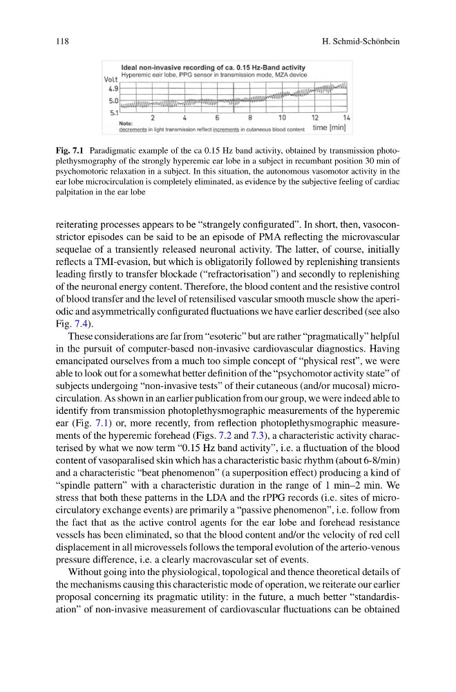
Fig. 7.1 Paradigmatic example of the ca 0.15 Hz band activity, obtained by transmission photoplethysmography of the strongly hyperemic ear lobe in a subject in recumbant position 30 min of
psychomotoric relaxation in a subject. In this situation, the autonomous vasomotor activity in the
ear lobe microcirculation is completely eliminated, as evidence by the subjective feeling of cardiac
palpitation in the ear lobe
reiterating processes appears to be “strangely configurated”. In short, then, vasoconstrictor episodes can be said to be an episode of PMA reflecting the microvascular
sequelae of a transiently released neuronal activity. The latter, of course, initially
reflects a TMI-evasion, but which is obligatorily followed by replenishing transients
leading firstly to transfer blockade (“refractorisation”) and secondly to replenishing
of the neuronal energy content. Therefore, the blood content and the resistive control
of blood transfer and the level of retensilised vascular smooth muscle show the aperiodic and asymmetrically configurated fluctuations we have earlier described (see also
Fig. 7.4).
These considerations are far from “esoteric” but are rather “pragmatically” helpful
in the pursuit of computer-based non-invasive cardiovascular diagnostics. Having
emancipated ourselves from a much too simple concept of “physical rest”, we were
able to look out for a somewhat better definition of the “psychomotor activity state” of
subjects undergoing “non-invasive tests” of their cutaneous (and/or mucosal) microcirculation. As shown in an earlier publication from our group, we were indeed able to
identify from transmission photoplethysmographic measurements of the hyperemic
ear (Fig. 7.1) or, more recently, from reflection photoplethysmographic measurements of the hyperemic forehead (Figs. 7.2 and 7.3), a characteristic activity characterised by what we now term “0.15 Hz band activity”, i.e. a fluctuation of the blood
content of vasoparalised skin which has a characteristic basic rhythm (about 6-8/min)
and a characteristic “beat phenomenon” (a superposition effect) producing a kind of
“spindle pattern” with a characteristic duration in the range of 1 min–2 min. We
stress that both these patterns in the LDA and the rPPG records (i.e. sites of microcirculatory exchange events) are primarily a “passive phenomenon”, i.e. follow from
the fact that as the active control agents for the ear lobe and forehead resistance
vessels has been eliminated, so that the blood content and/or the velocity of red cell
displacement in all microvessels follows the temporal evolution of the arterio-venous
pressure difference, i.e. a clearly macrovascular set of events.
Without going into the physiological, topological and thence theoretical details of
the mechanisms causing this characteristic mode of operation, we reiterate our earlier
proposal concerning its pragmatic utility: in the future, a much better “standardisation” of non-invasive measurement of cardiovascular fluctuations can be obtained
H. Schmid-Schönbein
Fig. 7.1 Paradigmatic example of the ca 0.15 Hz band activity, obtained by transmission photoplethysmography of the strongly hyperemic ear lobe in a subject in recumbant position 30 min of
psychomotoric relaxation in a subject. In this situation, the autonomous vasomotor activity in the
ear lobe microcirculation is completely eliminated, as evidence by the subjective feeling of cardiac
palpitation in the ear lobe
reiterating processes appears to be “strangely configurated”. In short, then, vasoconstrictor episodes can be said to be an episode of PMA reflecting the microvascular
sequelae of a transiently released neuronal activity. The latter, of course, initially
reflects a TMI-evasion, but which is obligatorily followed by replenishing transients
leading firstly to transfer blockade (“refractorisation”) and secondly to replenishing
of the neuronal energy content. Therefore, the blood content and the resistive control
of blood transfer and the level of retensilised vascular smooth muscle show the aperiodic and asymmetrically configurated fluctuations we have earlier described (see also
Fig. 7.4).
These considerations are far from “esoteric” but are rather “pragmatically” helpful
in the pursuit of computer-based non-invasive cardiovascular diagnostics. Having
emancipated ourselves from a much too simple concept of “physical rest”, we were
able to look out for a somewhat better definition of the “psychomotor activity state” of
subjects undergoing “non-invasive tests” of their cutaneous (and/or mucosal) microcirculation. As shown in an earlier publication from our group, we were indeed able to
identify from transmission photoplethysmographic measurements of the hyperemic
ear (Fig. 7.1) or, more recently, from reflection photoplethysmographic measurements of the hyperemic forehead (Figs. 7.2 and 7.3), a characteristic activity characterised by what we now term “0.15 Hz band activity”, i.e. a fluctuation of the blood
content of vasoparalised skin which has a characteristic basic rhythm (about 6-8/min)
and a characteristic “beat phenomenon” (a superposition effect) producing a kind of
“spindle pattern” with a characteristic duration in the range of 1 min–2 min. We
stress that both these patterns in the LDA and the rPPG records (i.e. sites of microcirculatory exchange events) are primarily a “passive phenomenon”, i.e. follow from
the fact that as the active control agents for the ear lobe and forehead resistance
vessels has been eliminated, so that the blood content and/or the velocity of red cell
displacement in all microvessels follows the temporal evolution of the arterio-venous
pressure difference, i.e. a clearly macrovascular set of events.
Without going into the physiological, topological and thence theoretical details of
the mechanisms causing this characteristic mode of operation, we reiterate our earlier
proposal concerning its pragmatic utility: in the future, a much better “standardisation” of non-invasive measurement of cardiovascular fluctuations can be obtained