

86
regulating vascular development [18]. Hyaluronan and its receptor, CD44, have
been shown to be essential in the formation and remodeling of blood vessels [18, 30,
62]. We have previously reported that a completely synthetic HA hydrogel can
maintain the self-renewal and pluripotency of hESCs [79, 86, 87]. Interestingly,
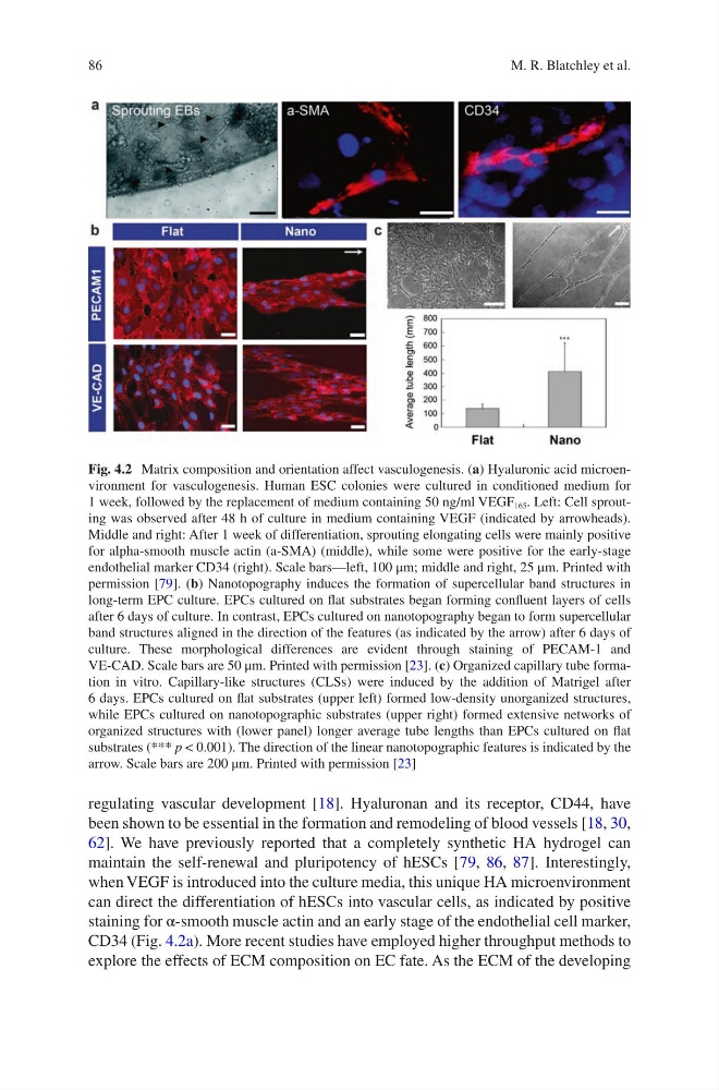
when VEGF is introduced into the culture media, this unique HA microenvironment
can direct the differentiation of hESCs into vascular cells, as indicated by positive
staining for α-smooth muscle actin and an early stage of the endothelial cell marker,
CD34 (Fig. 4.2a). More recent studies have employed higher throughput methods to
explore the effects of ECM composition on EC fate. As the ECM of the developing
Fig. 4.2 Matrix composition and orientation affect vasculogenesis. (a) Hyaluronic acid microenvironment for vasculogenesis. Human ESC colonies were cultured in conditioned medium for
1 week, followed by the replacement of medium containing 50 ng/ml VEGF 165 . Left: Cell sprouting was observed after 48 h of culture in medium containing VEGF (indicated by arrowheads).
Middle and right: After 1 week of differentiation, sprouting elongating cells were mainly positive
for alpha-smooth muscle actin (a-SMA) (middle), while some were positive for the early-stage
endothelial marker CD34 (right). Scale bars—left, 100 μm; middle and right, 25 μm. Printed with
permission [79]. (b) Nanotopography induces the formation of supercellular band structures in
long-term EPC culture. EPCs cultured on flat substrates began forming confluent layers of cells
after 6 days of culture. In contrast, EPCs cultured on nanotopography began to form supercellular
band structures aligned in the direction of the features (as indicated by the arrow) after 6 days of
culture. These morphological differences are evident through staining of PECAM-1 and
VE-CAD. Scale bars are 50 μm. Printed with permission [23]. (c) Organized capillary tube formation in vitro. Capillary-like structures (CLSs) were induced by the addition of Matrigel after
6 days. EPCs cultured on flat substrates (upper left) formed low-density unorganized structures,
while EPCs cultured on nanotopographic substrates (upper right) formed extensive networks of
organized structures with (lower panel) longer average tube lengths than EPCs cultured on flat
substrates (*** p < 0.001). The direction of the linear nanotopographic features is indicated by the
arrow. Scale bars are 200 μm. Printed with permission [23]
M. R. Blatchley et al.
regulating vascular development [18]. Hyaluronan and its receptor, CD44, have
been shown to be essential in the formation and remodeling of blood vessels [18, 30,
62]. We have previously reported that a completely synthetic HA hydrogel can
maintain the self-renewal and pluripotency of hESCs [79, 86, 87]. Interestingly,
when VEGF is introduced into the culture media, this unique HA microenvironment
can direct the differentiation of hESCs into vascular cells, as indicated by positive
staining for α-smooth muscle actin and an early stage of the endothelial cell marker,
CD34 (Fig. 4.2a). More recent studies have employed higher throughput methods to
explore the effects of ECM composition on EC fate. As the ECM of the developing
Fig. 4.2 Matrix composition and orientation affect vasculogenesis. (a) Hyaluronic acid microenvironment for vasculogenesis. Human ESC colonies were cultured in conditioned medium for
1 week, followed by the replacement of medium containing 50 ng/ml VEGF 165 . Left: Cell sprouting was observed after 48 h of culture in medium containing VEGF (indicated by arrowheads).
Middle and right: After 1 week of differentiation, sprouting elongating cells were mainly positive
for alpha-smooth muscle actin (a-SMA) (middle), while some were positive for the early-stage
endothelial marker CD34 (right). Scale bars—left, 100 μm; middle and right, 25 μm. Printed with
permission [79]. (b) Nanotopography induces the formation of supercellular band structures in
long-term EPC culture. EPCs cultured on flat substrates began forming confluent layers of cells
after 6 days of culture. In contrast, EPCs cultured on nanotopography began to form supercellular
band structures aligned in the direction of the features (as indicated by the arrow) after 6 days of
culture. These morphological differences are evident through staining of PECAM-1 and
VE-CAD. Scale bars are 50 μm. Printed with permission [23]. (c) Organized capillary tube formation in vitro. Capillary-like structures (CLSs) were induced by the addition of Matrigel after
6 days. EPCs cultured on flat substrates (upper left) formed low-density unorganized structures,
while EPCs cultured on nanotopographic substrates (upper right) formed extensive networks of
organized structures with (lower panel) longer average tube lengths than EPCs cultured on flat
substrates (*** p < 0.001). The direction of the linear nanotopographic features is indicated by the
arrow. Scale bars are 200 μm. Printed with permission [23]
M. R. Blatchley et al.