

42
scaffold and subjected to static strain aligned parallel to the strain direction [45].
These results agree with another report of ECs and muscle cells embedded within a
collagen-Matrigel 3D construct subjected to uniaxial stress [54]. In other work,
Chang et al. printed and framed aligned microvessel fragments embedded within a
collagen gel which was then subjected to uniaxial stress. While the pre-patterning
disappeared during culture in vitro, the vasculature within the constrained constructs aligned in parallel to the stretch direction, and both maintained alignment
during culture in vitro and induced invading vessels to align in the same direction
post-implantation [7].
The reasons for these recorded alignments are debatable. Ingber hypothesized
that local ECM thinning, caused by ECM turnover triggered by ECM modulators,
increases ECM compliance, resulting in the production of tractional forces by the
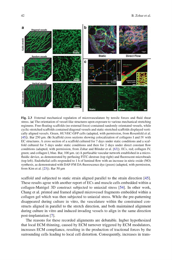
surrounding cells leading to local cell distortion. Consequently, increases in transFig. 2.3 External mechanical regulation of microvasculature by tensile forces and fluid shear
stress. (a) The orientation of vessel-like structures upon exposure to various mechanical stretching
regimens. Free-floating scaffolds (no external force) contained randomly orientated vessels, while
cyclic-stretched scaffolds contained diagonal vessels and static-stretched scaffolds displayed vertically aligned vessels. Green, HUVEC-GFP cells (adapted, with permission, from Rosenfeld et al.
[45]). Bar 250 μm. (b) Scaffold cross sections showing colocalization of collagens I and IV with
EC structures. A cross section of a scaffold cultured for 7 days under static conditions and a scaffold cultured for 5 days under static conditions and then for 2 days under direct constant flow
conditions (adapted, with permission, from Zohar and Blinder et al. [63]). ECs, red; collagen IV,
green; and collagen I, blue. Bar, 100 μm. (c) A perfusable vascular network established in a microfluidic device, as demonstrated by perfusing FITC-dextran (top right) and fluorescent microbeads
(top left). Endothelial cells responded to 1 h of luminal flow with an increase in nitric oxide (NO)
synthesis, as demonstrated with DAF-FM DA fluorescence dye (green) (adapted, with permission,
from Kim et al. [23]). Bar 50 μm
B. Zohar et al.
scaffold and subjected to static strain aligned parallel to the strain direction [45].
These results agree with another report of ECs and muscle cells embedded within a
collagen-Matrigel 3D construct subjected to uniaxial stress [54]. In other work,
Chang et al. printed and framed aligned microvessel fragments embedded within a
collagen gel which was then subjected to uniaxial stress. While the pre-patterning
disappeared during culture in vitro, the vasculature within the constrained constructs aligned in parallel to the stretch direction, and both maintained alignment
during culture in vitro and induced invading vessels to align in the same direction
post-implantation [7].
The reasons for these recorded alignments are debatable. Ingber hypothesized
that local ECM thinning, caused by ECM turnover triggered by ECM modulators,
increases ECM compliance, resulting in the production of tractional forces by the
surrounding cells leading to local cell distortion. Consequently, increases in transFig. 2.3 External mechanical regulation of microvasculature by tensile forces and fluid shear
stress. (a) The orientation of vessel-like structures upon exposure to various mechanical stretching
regimens. Free-floating scaffolds (no external force) contained randomly orientated vessels, while
cyclic-stretched scaffolds contained diagonal vessels and static-stretched scaffolds displayed vertically aligned vessels. Green, HUVEC-GFP cells (adapted, with permission, from Rosenfeld et al.
[45]). Bar 250 μm. (b) Scaffold cross sections showing colocalization of collagens I and IV with
EC structures. A cross section of a scaffold cultured for 7 days under static conditions and a scaffold cultured for 5 days under static conditions and then for 2 days under direct constant flow
conditions (adapted, with permission, from Zohar and Blinder et al. [63]). ECs, red; collagen IV,
green; and collagen I, blue. Bar, 100 μm. (c) A perfusable vascular network established in a microfluidic device, as demonstrated by perfusing FITC-dextran (top right) and fluorescent microbeads
(top left). Endothelial cells responded to 1 h of luminal flow with an increase in nitric oxide (NO)
synthesis, as demonstrated with DAF-FM DA fluorescence dye (green) (adapted, with permission,
from Kim et al. [23]). Bar 50 μm
B. Zohar et al.