

22
of PDGF-BB and HB-EGF [96]. Blockade of these growth factors using blocking
antibodies, receptor traps, or chemical disruption of their receptors revealed their
involvement [96]. Importantly, we assessed pericyte motility and invasion under
our serum-free media conditions and demonstrated that they failed to move, when
cultured by themselves. In contrast, when pericytes were cocultured with ECs, pericyte motility ensued allowing for recruitment to EC-lined tubes [96]. This
EC-dependent pericyte motility response was strongly abrogated by the combined
blocking reagents directed at both PDGF-BB and HB-EGF [96]. These findings are
consistent with work showing that EC-specific knockout of PDGF-BB leads to
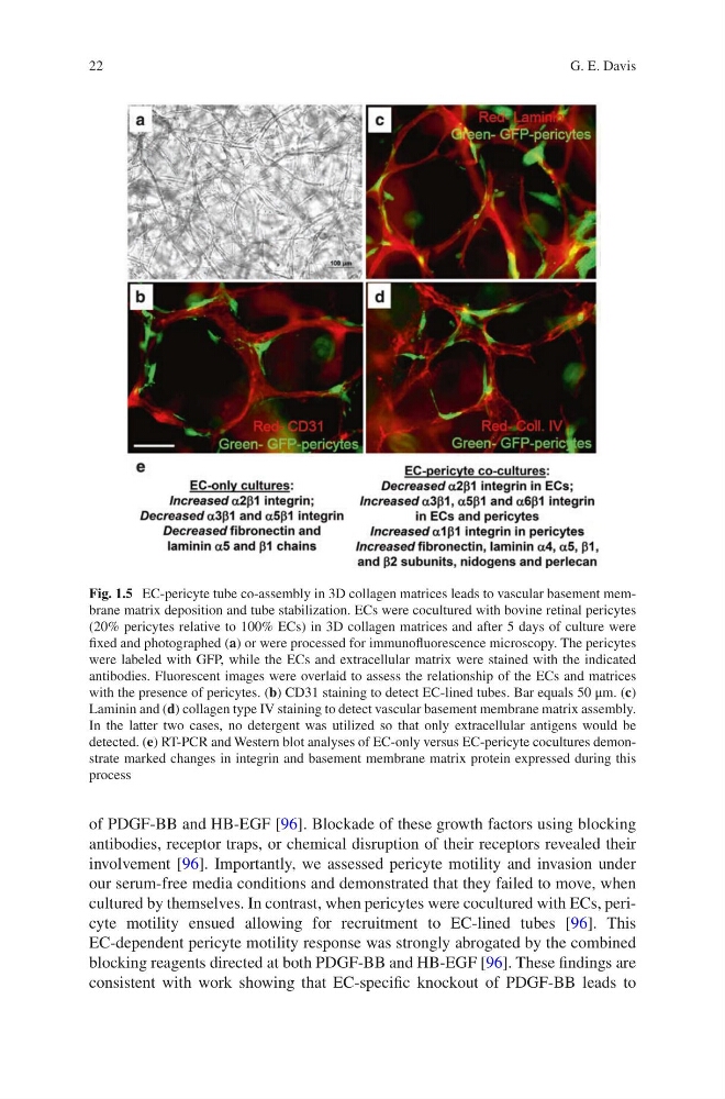
Fig. 1.5 EC-pericyte tube co-assembly in 3D collagen matrices leads to vascular basement membrane matrix deposition and tube stabilization. ECs were cocultured with bovine retinal pericytes
(20% pericytes relative to 100% ECs) in 3D collagen matrices and after 5 days of culture were
fixed and photographed (a) or were processed for immunofluorescence microscopy. The pericytes
were labeled with GFP, while the ECs and extracellular matrix were stained with the indicated
antibodies. Fluorescent images were overlaid to assess the relationship of the ECs and matrices
with the presence of pericytes. (b) CD31 staining to detect EC-lined tubes. Bar equals 50 μm. (c)
Laminin and (d) collagen type IV staining to detect vascular basement membrane matrix assembly.
In the latter two cases, no detergent was utilized so that only extracellular antigens would be
detected. (e) RT-PCR and Western blot analyses of EC-only versus EC-pericyte cocultures demonstrate marked changes in integrin and basement membrane matrix protein expressed during this
process
G. E. Davis
of PDGF-BB and HB-EGF [96]. Blockade of these growth factors using blocking
antibodies, receptor traps, or chemical disruption of their receptors revealed their
involvement [96]. Importantly, we assessed pericyte motility and invasion under
our serum-free media conditions and demonstrated that they failed to move, when
cultured by themselves. In contrast, when pericytes were cocultured with ECs, pericyte motility ensued allowing for recruitment to EC-lined tubes [96]. This
EC-dependent pericyte motility response was strongly abrogated by the combined
blocking reagents directed at both PDGF-BB and HB-EGF [96]. These findings are
consistent with work showing that EC-specific knockout of PDGF-BB leads to
Fig. 1.5 EC-pericyte tube co-assembly in 3D collagen matrices leads to vascular basement membrane matrix deposition and tube stabilization. ECs were cocultured with bovine retinal pericytes
(20% pericytes relative to 100% ECs) in 3D collagen matrices and after 5 days of culture were
fixed and photographed (a) or were processed for immunofluorescence microscopy. The pericytes
were labeled with GFP, while the ECs and extracellular matrix were stained with the indicated
antibodies. Fluorescent images were overlaid to assess the relationship of the ECs and matrices
with the presence of pericytes. (b) CD31 staining to detect EC-lined tubes. Bar equals 50 μm. (c)
Laminin and (d) collagen type IV staining to detect vascular basement membrane matrix assembly.
In the latter two cases, no detergent was utilized so that only extracellular antigens would be
detected. (e) RT-PCR and Western blot analyses of EC-only versus EC-pericyte cocultures demonstrate marked changes in integrin and basement membrane matrix protein expressed during this
process
G. E. Davis