

14
TIMP-4 have dramatic blocking effects on both sprouting and lumen formation is
more supportive of a major role for MT-MMPs, rather than ADAMs, since there is
currently no evidence to suggest that they can block ADAM proteinases [7].
A further important experiment which demonstrates a role for MT1-MMP during EC tubulogenesis is that increased expression of MT1-MMP using viral vectors
leads to marked increases in lumen formation that depends on its MMP catalytic
domain [95]. Furthermore, addition of GM6001 to block MT1-MMP completely
inhibits the stimulatory influence of the recombinant protein. In support of this
result is that a catalytically dead full-length MT1-MMP construct (EA mutant) has
no ability to stimulate EC lumen formation [95], while interestingly, it does not
exert an inhibitory influence. In very recent experiments, we have further shown that
increasing the expression of wild-type, full-length MT1-MMP increases both the
rate and extent of EC lumen formation in 3D collagen matrices [83]. Of great interest is that we have created a construct that appears to be a dominant-negative mutant
of MT1-MMP in this system where we mutated the active site and at the same time
deleted its cytoplasmic tail [83]. When expressed in ECs, the cells are completely
unable to make lumens in 3D collagen matrices. An additional finding is that
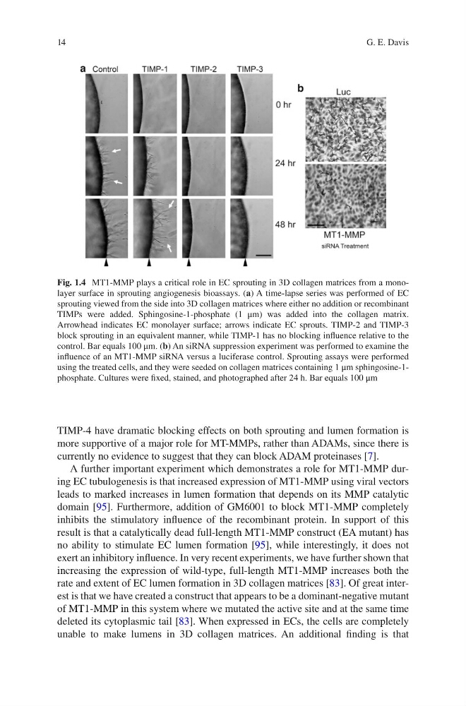
Fig. 1.4 MT1-MMP plays a critical role in EC sprouting in 3D collagen matrices from a monolayer surface in sprouting angiogenesis bioassays. (a) A time-lapse series was performed of EC
sprouting viewed from the side into 3D collagen matrices where either no addition or recombinant
TIMPs were added. Sphingosine-1-phosphate (1 μm) was added into the collagen matrix.
Arrowhead indicates EC monolayer surface; arrows indicate EC sprouts. TIMP-2 and TIMP-3
block sprouting in an equivalent manner, while TIMP-1 has no blocking influence relative to the
control. Bar equals 100 μm. (b) An siRNA suppression experiment was performed to examine the
influence of an MT1-MMP siRNA versus a luciferase control. Sprouting assays were performed
using the treated cells, and they were seeded on collagen matrices containing 1 μm sphingosine- 1phosphate. Cultures were fixed, stained, and photographed after 24 h. Bar equals 100 μm
G. E. Davis
TIMP-4 have dramatic blocking effects on both sprouting and lumen formation is
more supportive of a major role for MT-MMPs, rather than ADAMs, since there is
currently no evidence to suggest that they can block ADAM proteinases [7].
A further important experiment which demonstrates a role for MT1-MMP during EC tubulogenesis is that increased expression of MT1-MMP using viral vectors
leads to marked increases in lumen formation that depends on its MMP catalytic
domain [95]. Furthermore, addition of GM6001 to block MT1-MMP completely
inhibits the stimulatory influence of the recombinant protein. In support of this
result is that a catalytically dead full-length MT1-MMP construct (EA mutant) has
no ability to stimulate EC lumen formation [95], while interestingly, it does not
exert an inhibitory influence. In very recent experiments, we have further shown that
increasing the expression of wild-type, full-length MT1-MMP increases both the
rate and extent of EC lumen formation in 3D collagen matrices [83]. Of great interest is that we have created a construct that appears to be a dominant-negative mutant
of MT1-MMP in this system where we mutated the active site and at the same time
deleted its cytoplasmic tail [83]. When expressed in ECs, the cells are completely
unable to make lumens in 3D collagen matrices. An additional finding is that
Fig. 1.4 MT1-MMP plays a critical role in EC sprouting in 3D collagen matrices from a monolayer surface in sprouting angiogenesis bioassays. (a) A time-lapse series was performed of EC
sprouting viewed from the side into 3D collagen matrices where either no addition or recombinant
TIMPs were added. Sphingosine-1-phosphate (1 μm) was added into the collagen matrix.
Arrowhead indicates EC monolayer surface; arrows indicate EC sprouts. TIMP-2 and TIMP-3
block sprouting in an equivalent manner, while TIMP-1 has no blocking influence relative to the
control. Bar equals 100 μm. (b) An siRNA suppression experiment was performed to examine the
influence of an MT1-MMP siRNA versus a luciferase control. Sprouting assays were performed
using the treated cells, and they were seeded on collagen matrices containing 1 μm sphingosine- 1phosphate. Cultures were fixed, stained, and photographed after 24 h. Bar equals 100 μm
G. E. Davis