

10
1.3.4 Identification of New Small GTPases and Their Effectors
Controlling EC Tube Morphogenesis
Recent studies have sought to identify additional small GTPases and downstream
effectors that regulate the process of capillary tube morphogenesis. We demonstrated critical roles for k-Ras, Rac2, and Rap1b as well as a new series of effectors
including Rasip1, IQGAP1, MRCKβ, GIT1, and beta-Pix (Fig. 1.2) [79]. siRNA
suppression of these molecules individually or in combination resulted in marked
defects in EC lumen and tube assembly [79]. In contrast, as in past studies, siRNA
suppression of RhoA did not interfere with these processes. Interestingly, Rasip1 is
known to interact with the RhoA-specific GTPase-activating protein (GAP),
Arhgap29 [102], and siRNA suppression of this GAP lead to reduced lumen formation because of elevated RhoA activity [79]. Importantly, we also identified two
additional GAPs that negatively regulate EC lumen formation, which are Arhgap31
(which inactivates Cdc42 and Rac1) and Rasa1 (which inactivates Ras isoforms)
(Fig. 1.2). Combined siRNA suppression of Arhgap31 and Rasa1 led to marked
increases in EC tube formation, suggesting an important combined role for Cdc42/
Rac/k-Ras and possibly other Ras isoforms [79]. Combined siRNA suppression of
Cdc42 with k-Ras leads to strong blockade of EC lumen and tube assembly [79].
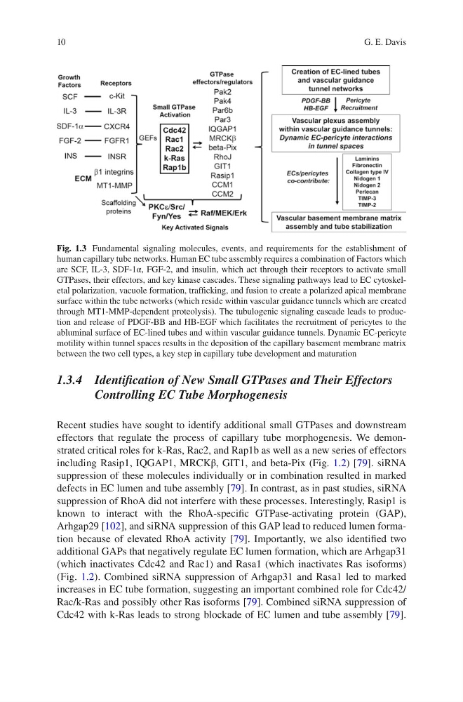
Fig. 1.3 Fundamental signaling molecules, events, and requirements for the establishment of
human capillary tube networks. Human EC tube assembly requires a combination of Factors which
are SCF, IL-3, SDF-1α, FGF-2, and insulin, which act through their receptors to activate small
GTPases, their effectors, and key kinase cascades. These signaling pathways lead to EC cytoskeletal polarization, vacuole formation, trafficking, and fusion to create a polarized apical membrane
surface within the tube networks (which reside within vascular guidance tunnels which are created
through MT1-MMP-dependent proteolysis). The tubulogenic signaling cascade leads to production and release of PDGF-BB and HB-EGF which facilitates the recruitment of pericytes to the
abluminal surface of EC-lined tubes and within vascular guidance tunnels. Dynamic EC-pericyte
motility within tunnel spaces results in the deposition of the capillary basement membrane matrix
between the two cell types, a key step in capillary tube development and maturation
G. E. Davis
1.3.4 Identification of New Small GTPases and Their Effectors
Controlling EC Tube Morphogenesis
Recent studies have sought to identify additional small GTPases and downstream
effectors that regulate the process of capillary tube morphogenesis. We demonstrated critical roles for k-Ras, Rac2, and Rap1b as well as a new series of effectors
including Rasip1, IQGAP1, MRCKβ, GIT1, and beta-Pix (Fig. 1.2) [79]. siRNA
suppression of these molecules individually or in combination resulted in marked
defects in EC lumen and tube assembly [79]. In contrast, as in past studies, siRNA
suppression of RhoA did not interfere with these processes. Interestingly, Rasip1 is
known to interact with the RhoA-specific GTPase-activating protein (GAP),
Arhgap29 [102], and siRNA suppression of this GAP lead to reduced lumen formation because of elevated RhoA activity [79]. Importantly, we also identified two
additional GAPs that negatively regulate EC lumen formation, which are Arhgap31
(which inactivates Cdc42 and Rac1) and Rasa1 (which inactivates Ras isoforms)
(Fig. 1.2). Combined siRNA suppression of Arhgap31 and Rasa1 led to marked
increases in EC tube formation, suggesting an important combined role for Cdc42/
Rac/k-Ras and possibly other Ras isoforms [79]. Combined siRNA suppression of
Cdc42 with k-Ras leads to strong blockade of EC lumen and tube assembly [79].
Fig. 1.3 Fundamental signaling molecules, events, and requirements for the establishment of
human capillary tube networks. Human EC tube assembly requires a combination of Factors which
are SCF, IL-3, SDF-1α, FGF-2, and insulin, which act through their receptors to activate small
GTPases, their effectors, and key kinase cascades. These signaling pathways lead to EC cytoskeletal polarization, vacuole formation, trafficking, and fusion to create a polarized apical membrane
surface within the tube networks (which reside within vascular guidance tunnels which are created
through MT1-MMP-dependent proteolysis). The tubulogenic signaling cascade leads to production and release of PDGF-BB and HB-EGF which facilitates the recruitment of pericytes to the
abluminal surface of EC-lined tubes and within vascular guidance tunnels. Dynamic EC-pericyte
motility within tunnel spaces results in the deposition of the capillary basement membrane matrix
between the two cell types, a key step in capillary tube development and maturation
G. E. Davis