

181
during and after brain transplantation, (2) the success of a therapy is widely dependent on the interaction between the transplanted/injected material and the host tissue, (3) the material degradation rate and composition must be finely tuned to allow
tissue growth, and (4) the success of a pro-repair therapy relies on the simultaneous
activation of axonal sprouting, vascular growth, neurogenesis, and modulation of
the injury-induced inflammation, each presenting unique biological challenges.
In recent years, several experimental studies developed injectable hydrogels and
growth factor delivery systems to promote poststroke angiogenesis through VEGF
administration. A 2010 study from Emerich D.F. et al. describes the effect of an
injectable VEGF-loaded alginate-based hydrogel on poststroke functional recovery
[11]. Behavioral testing shows that the performance of rats, receiving VEGF gels in
the striatum 15 min prior to stroke, was significantly improved relative to a blank
gel with or without VEGF. This study strengthens previous evidence that VEGF
exerts neuroprotective properties when upregulated prior to injury.
Zhang et al. attempt to restore tissue integrity in a rat model of brain damage by
implanting a porous polydimethylsiloxane-tetraethoxysilane (PDMS-TEOS) hybrid
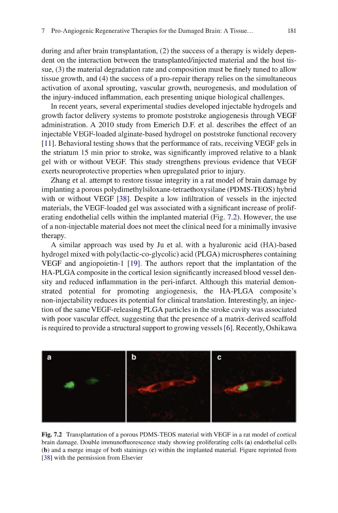
with or without VEGF [38]. Despite a low infiltration of vessels in the injected
materials, the VEGF-loaded gel was associated with a significant increase of proliferating endothelial cells within the implanted material (Fig. 7.2). However, the use
of a non-injectable material does not meet the clinical need for a minimally invasive
therapy.
A similar approach was used by Ju et al. with a hyaluronic acid (HA)-based
hydrogel mixed with poly(lactic-co-glycolic) acid (PLGA) microspheres containing
VEGF and angiopoietin-1 [19]. The authors report that the implantation of the
HA-PLGA composite in the cortical lesion significantly increased blood vessel density and reduced inflammation in the peri-infarct. Although this material demonstrated potential for promoting angiogenesis, the HA-PLGA composite’s
non-injectability reduces its potential for clinical translation. Interestingly, an injection of the same VEGF-releasing PLGA particles in the stroke cavity was associated
with poor vascular effect, suggesting that the presence of a matrix-derived scaffold
is required to provide a structural support to growing vessels [6]. Recently, Oshikawa
Fig. 7.2 Transplantation of a porous PDMS-TEOS material with VEGF in a rat model of cortical
brain damage. Double immunofluorescence study showing proliferating cells (a) endothelial cells
(b) and a merge image of both stainings (c) within the implanted material. Figure reprinted from
[38] with the permission from Elsevier
7 Pro-Angiogenic Regenerative Therapies for the Damaged Brain: A Tissue…
during and after brain transplantation, (2) the success of a therapy is widely dependent on the interaction between the transplanted/injected material and the host tissue, (3) the material degradation rate and composition must be finely tuned to allow
tissue growth, and (4) the success of a pro-repair therapy relies on the simultaneous
activation of axonal sprouting, vascular growth, neurogenesis, and modulation of
the injury-induced inflammation, each presenting unique biological challenges.
In recent years, several experimental studies developed injectable hydrogels and
growth factor delivery systems to promote poststroke angiogenesis through VEGF
administration. A 2010 study from Emerich D.F. et al. describes the effect of an
injectable VEGF-loaded alginate-based hydrogel on poststroke functional recovery
[11]. Behavioral testing shows that the performance of rats, receiving VEGF gels in
the striatum 15 min prior to stroke, was significantly improved relative to a blank
gel with or without VEGF. This study strengthens previous evidence that VEGF
exerts neuroprotective properties when upregulated prior to injury.
Zhang et al. attempt to restore tissue integrity in a rat model of brain damage by
implanting a porous polydimethylsiloxane-tetraethoxysilane (PDMS-TEOS) hybrid
with or without VEGF [38]. Despite a low infiltration of vessels in the injected
materials, the VEGF-loaded gel was associated with a significant increase of proliferating endothelial cells within the implanted material (Fig. 7.2). However, the use
of a non-injectable material does not meet the clinical need for a minimally invasive
therapy.
A similar approach was used by Ju et al. with a hyaluronic acid (HA)-based
hydrogel mixed with poly(lactic-co-glycolic) acid (PLGA) microspheres containing
VEGF and angiopoietin-1 [19]. The authors report that the implantation of the
HA-PLGA composite in the cortical lesion significantly increased blood vessel density and reduced inflammation in the peri-infarct. Although this material demonstrated potential for promoting angiogenesis, the HA-PLGA composite’s
non-injectability reduces its potential for clinical translation. Interestingly, an injection of the same VEGF-releasing PLGA particles in the stroke cavity was associated
with poor vascular effect, suggesting that the presence of a matrix-derived scaffold
is required to provide a structural support to growing vessels [6]. Recently, Oshikawa
Fig. 7.2 Transplantation of a porous PDMS-TEOS material with VEGF in a rat model of cortical
brain damage. Double immunofluorescence study showing proliferating cells (a) endothelial cells
(b) and a merge image of both stainings (c) within the implanted material. Figure reprinted from
[38] with the permission from Elsevier
7 Pro-Angiogenic Regenerative Therapies for the Damaged Brain: A Tissue…