

179
suggests that newly formed vessels in the periphery of the injured site mediate
recovery via the activation of endogenous mechanisms of plasticity [7]. In particular, it was shown that angiogenesis and neurogenesis not only work in tandem but
are also causally linked. The daily administration of an angiogenesis inhibitor, in a
mouse model of ischemic stroke, is associated with a significant reduction of proliferation, migration, and differentiation of endogenous neural progenitors which
impairs their functional recovery [24, 26]. In addition, aggregate data supports a
“cleanup hypothesis,” whereby the newly formed vessels in the penumbra serve to
facilitate macrophage infiltration and clear up necrotic debris [22, 37].
7.4 Role of VEGF in Endogenous Angiogenesis
In both experimental and clinical studies, enhanced vessel formation and restored
perfusion in the ischemic border correlate with improved long-term recovery and
longer survival times [20]. Older patients, who tend to do worse after stroke, often
show reduced new vessel formation in the penumbra [2, 12, 34]. Similarly, patients
who develop dementia after stroke have reduced blood flow in cortical regions adjacent to the stroke [30]. Pro-repair factors correlated with brain angiogenesis have
also been extensively assessed in experimental stroke models. One major endogenous signal, vascular endothelial growth factor (VEGF) is found in both neurons
and astrocytes after cerebral ischemia [1, 39]. A positive correlation was found
between the severity of damages and the concentration of VEGF in stroke patients
[31]. Likewise, postmortem studies reveal an increased expression of the proangiogenic factor and its receptor VEGFR-2, both at mRNA and protein levels in
the penumbra [17]. In addition, studies have shown that a deficit in VEGF distribution is associated with disorganized and impaired vessel formation, suggesting that
ECM-binding VEGF isoforms provide essential stimulatory cues to initiate vessel
branching [29].
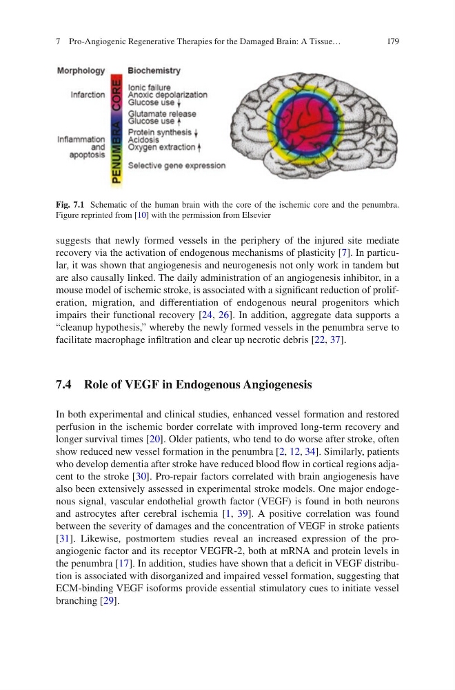
Fig. 7.1 Schematic of the human brain with the core of the ischemic core and the penumbra.
Figure reprinted from [10] with the permission from Elsevier
7 Pro-Angiogenic Regenerative Therapies for the Damaged Brain: A Tissue…
suggests that newly formed vessels in the periphery of the injured site mediate
recovery via the activation of endogenous mechanisms of plasticity [7]. In particular, it was shown that angiogenesis and neurogenesis not only work in tandem but
are also causally linked. The daily administration of an angiogenesis inhibitor, in a
mouse model of ischemic stroke, is associated with a significant reduction of proliferation, migration, and differentiation of endogenous neural progenitors which
impairs their functional recovery [24, 26]. In addition, aggregate data supports a
“cleanup hypothesis,” whereby the newly formed vessels in the penumbra serve to
facilitate macrophage infiltration and clear up necrotic debris [22, 37].
7.4 Role of VEGF in Endogenous Angiogenesis
In both experimental and clinical studies, enhanced vessel formation and restored
perfusion in the ischemic border correlate with improved long-term recovery and
longer survival times [20]. Older patients, who tend to do worse after stroke, often
show reduced new vessel formation in the penumbra [2, 12, 34]. Similarly, patients
who develop dementia after stroke have reduced blood flow in cortical regions adjacent to the stroke [30]. Pro-repair factors correlated with brain angiogenesis have
also been extensively assessed in experimental stroke models. One major endogenous signal, vascular endothelial growth factor (VEGF) is found in both neurons
and astrocytes after cerebral ischemia [1, 39]. A positive correlation was found
between the severity of damages and the concentration of VEGF in stroke patients
[31]. Likewise, postmortem studies reveal an increased expression of the proangiogenic factor and its receptor VEGFR-2, both at mRNA and protein levels in
the penumbra [17]. In addition, studies have shown that a deficit in VEGF distribution is associated with disorganized and impaired vessel formation, suggesting that
ECM-binding VEGF isoforms provide essential stimulatory cues to initiate vessel
branching [29].
Fig. 7.1 Schematic of the human brain with the core of the ischemic core and the penumbra.
Figure reprinted from [10] with the permission from Elsevier
7 Pro-Angiogenic Regenerative Therapies for the Damaged Brain: A Tissue…