

6
intermixing of cytoplasmic contents when this occurs, so the tube consists of adjacent ECs which interact through cell-cell adhesive contacts. In addition, the ECs are
attached to the ECM to form the wall of a luminal space and need to maintain these
adhesive contacts (i.e., both cell-ECM and cell-cell adhesion) to remain stable on
this luminal wall. Intracellular vacuoles can be observed to form through integrinand cytoskeletal-dependent pinocytic events, and these vacuoles target to a pericentrosomal location in a polarized fashion and then move to fuse with the developing
luminal membrane as shown in Fig. 1.1b (upper panel) [8, 28, 30, 62–64, 104]. In
Fig. 1.1b, intracellular vacuoles are strongly labeled with a GFP-Rac1 construct [8].
We previously observed labeling of intracellular vacuoles with GFP-Rac1, GFPCdc42, and GFP-RalA [8, 30, 79]. This GFP-Cdc42 was expressed using an
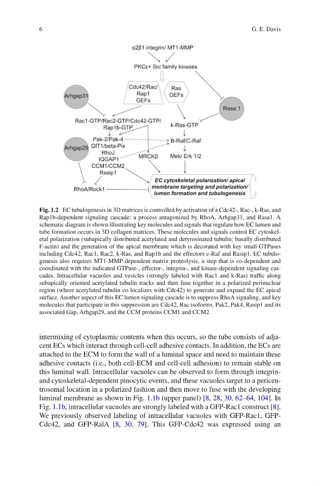
Fig. 1.2 EC tubulogenesis in 3D matrices is controlled by activation of a Cdc42-, Rac-, k-Ras, and
Rap1b-dependent signaling cascade: a process antagonized by RhoA, Arhgap31, and Rasa1. A
schematic diagram is shown illustrating key molecules and signals that regulate how EC lumen and
tube formation occurs in 3D collagen matrices. These molecules and signals control EC cytoskeletal polarization (subapically distributed acetylated and detyrosinated tubulin; basally distributed
F-actin) and the generation of the apical membrane which is decorated with key small GTPases
including Cdc42, Rac1, Rac2, k-Ras, and Rap1b and the effectors c-Raf and Rasip1. EC tubulogenesis also requires MT1-MMP-dependent matrix proteolysis, a step that is co-dependent and
coordinated with the indicated GTPase-, effector-, integrin-, and kinase-dependent signaling cascades. Intracellular vacuoles and vesicles (strongly labeled with Rac1 and k-Ras) traffic along
subapically oriented acetylated tubulin tracks and then fuse together in a polarized perinuclear
region (where acetylated tubulin co-localizes with Cdc42) to generate and expand the EC apical
surface. Another aspect of this EC lumen signaling cascade is to suppress RhoA signaling, and key
molecules that participate in this suppression are Cdc42, Rac isoforms, Pak2, Pak4, Rasip1 and its
associated Gap, Arhgap29, and the CCM proteins CCM1 and CCM2
G. E. Davis
intermixing of cytoplasmic contents when this occurs, so the tube consists of adjacent ECs which interact through cell-cell adhesive contacts. In addition, the ECs are
attached to the ECM to form the wall of a luminal space and need to maintain these
adhesive contacts (i.e., both cell-ECM and cell-cell adhesion) to remain stable on
this luminal wall. Intracellular vacuoles can be observed to form through integrinand cytoskeletal-dependent pinocytic events, and these vacuoles target to a pericentrosomal location in a polarized fashion and then move to fuse with the developing
luminal membrane as shown in Fig. 1.1b (upper panel) [8, 28, 30, 62–64, 104]. In
Fig. 1.1b, intracellular vacuoles are strongly labeled with a GFP-Rac1 construct [8].
We previously observed labeling of intracellular vacuoles with GFP-Rac1, GFPCdc42, and GFP-RalA [8, 30, 79]. This GFP-Cdc42 was expressed using an
Fig. 1.2 EC tubulogenesis in 3D matrices is controlled by activation of a Cdc42-, Rac-, k-Ras, and
Rap1b-dependent signaling cascade: a process antagonized by RhoA, Arhgap31, and Rasa1. A
schematic diagram is shown illustrating key molecules and signals that regulate how EC lumen and
tube formation occurs in 3D collagen matrices. These molecules and signals control EC cytoskeletal polarization (subapically distributed acetylated and detyrosinated tubulin; basally distributed
F-actin) and the generation of the apical membrane which is decorated with key small GTPases
including Cdc42, Rac1, Rac2, k-Ras, and Rap1b and the effectors c-Raf and Rasip1. EC tubulogenesis also requires MT1-MMP-dependent matrix proteolysis, a step that is co-dependent and
coordinated with the indicated GTPase-, effector-, integrin-, and kinase-dependent signaling cascades. Intracellular vacuoles and vesicles (strongly labeled with Rac1 and k-Ras) traffic along
subapically oriented acetylated tubulin tracks and then fuse together in a polarized perinuclear
region (where acetylated tubulin co-localizes with Cdc42) to generate and expand the EC apical
surface. Another aspect of this EC lumen signaling cascade is to suppress RhoA signaling, and key
molecules that participate in this suppression are Cdc42, Rac isoforms, Pak2, Pak4, Rasip1 and its
associated Gap, Arhgap29, and the CCM proteins CCM1 and CCM2
G. E. Davis