

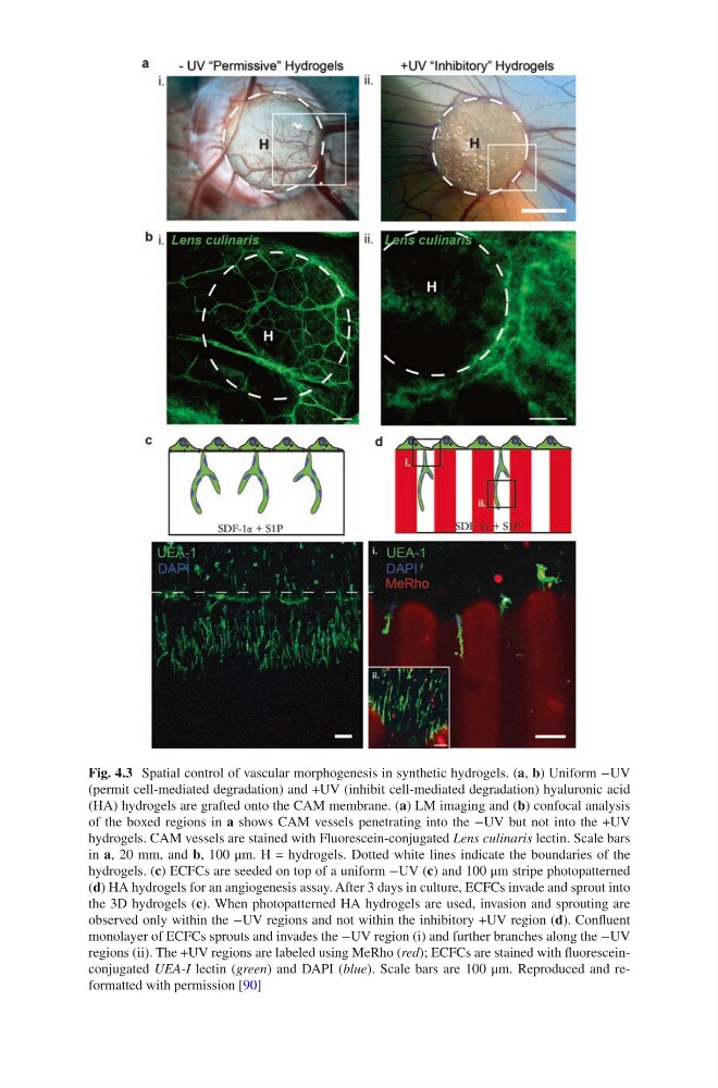
Fig. 4.3 Spatial control of vascular morphogenesis in synthetic hydrogels. (a, b) Uniform −UV
(permit cell-mediated degradation) and +UV (inhibit cell-mediated degradation) hyaluronic acid
(HA) hydrogels are grafted onto the CAM membrane. (a) LM imaging and (b) confocal analysis
of the boxed regions in a shows CAM vessels penetrating into the −UV but not into the +UV
hydrogels. CAM vessels are stained with Fluorescein-conjugated Lens culinaris lectin. Scale bars
in a, 20 mm, and b, 100 μm. H = hydrogels. Dotted white lines indicate the boundaries of the
hydrogels. (c) ECFCs are seeded on top of a uniform −UV (c) and 100 μm stripe photopatterned
(d) HA hydrogels for an angiogenesis assay. After 3 days in culture, ECFCs invade and sprout into
the 3D hydrogels (c). When photopatterned HA hydrogels are used, invasion and sprouting are
observed only within the −UV regions and not within the inhibitory +UV region (d). Confluent
monolayer of ECFCs sprouts and invades the −UV region (i) and further branches along the −UV
regions (ii). The +UV regions are labeled using MeRho (red); ECFCs are stained with fluoresceinconjugated UEA-I lectin (green) and DAPI (blue). Scale bars are 100 μm. Reproduced and reformatted with permission [90]
(permit cell-mediated degradation) and +UV (inhibit cell-mediated degradation) hyaluronic acid
(HA) hydrogels are grafted onto the CAM membrane. (a) LM imaging and (b) confocal analysis
of the boxed regions in a shows CAM vessels penetrating into the −UV but not into the +UV
hydrogels. CAM vessels are stained with Fluorescein-conjugated Lens culinaris lectin. Scale bars
in a, 20 mm, and b, 100 μm. H = hydrogels. Dotted white lines indicate the boundaries of the
hydrogels. (c) ECFCs are seeded on top of a uniform −UV (c) and 100 μm stripe photopatterned
(d) HA hydrogels for an angiogenesis assay. After 3 days in culture, ECFCs invade and sprout into
the 3D hydrogels (c). When photopatterned HA hydrogels are used, invasion and sprouting are
observed only within the −UV regions and not within the inhibitory +UV region (d). Confluent
monolayer of ECFCs sprouts and invades the −UV region (i) and further branches along the −UV
regions (ii). The +UV regions are labeled using MeRho (red); ECFCs are stained with fluoresceinconjugated UEA-I lectin (green) and DAPI (blue). Scale bars are 100 μm. Reproduced and reformatted with permission [90]