

DF image when the outer collection angle is not sufficiently large. In such a case, a
considerable fraction of the scattering ends up beyond the DF detector, e.g. when
the scattering distribution broadens upon increase of thickness with specimen tilt.
Then the contrast of inclusions in a thick matrix, such as clusters of stain reagents,
could be inverted. These effects are even more pronounced when energy-selected
images are used to amplify the effect of inelastic scattering from the lighter matrix
with respect to the scattering of a heavily stained reagent [7].
2.2.4 Elemental Analysis
The ability to perform both multimodal imaging and spectroscopy makes STEM a
truly powerful analytical technique for studying biological materials [8, 9, 19].
DF STEM collects electrons scattered at pre-defined high angles for image formation, while simultaneously allowing small-angle scattered electrons to pass
through the opening in the detector into an electron energy-loss spectrometer
(EELS). STEM is ideally suited for elemental analysis of biological samples when
beam damage is the limiting factor, since the entire spectrum is collected in parallel.
This is in contrast to energy filtered TEM where images from successive
energy-loss windows are recorded sequentially. In STEM, the electron dose is
applied only to the volume that is analyzed, and the dose rate can be controlled
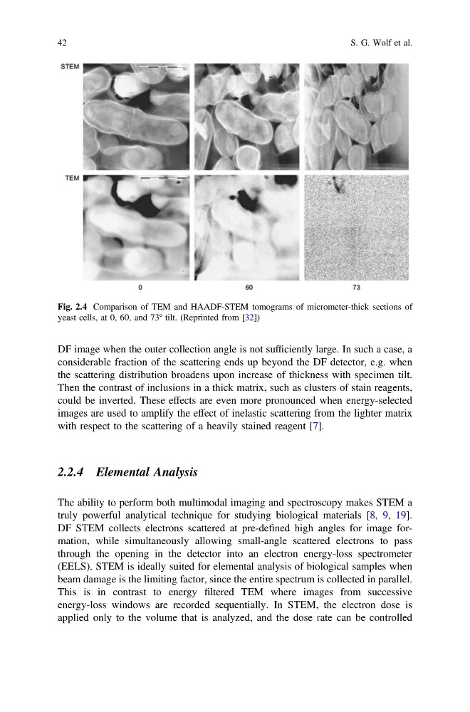
Fig. 2.4 Comparison of TEM and HAADF-STEM tomograms of micrometer-thick sections of
yeast cells, at 0, 60, and 73º tilt. (Reprinted from [32])
42
S. G. Wolf et al.
considerable fraction of the scattering ends up beyond the DF detector, e.g. when
the scattering distribution broadens upon increase of thickness with specimen tilt.
Then the contrast of inclusions in a thick matrix, such as clusters of stain reagents,
could be inverted. These effects are even more pronounced when energy-selected
images are used to amplify the effect of inelastic scattering from the lighter matrix
with respect to the scattering of a heavily stained reagent [7].
2.2.4 Elemental Analysis
The ability to perform both multimodal imaging and spectroscopy makes STEM a
truly powerful analytical technique for studying biological materials [8, 9, 19].
DF STEM collects electrons scattered at pre-defined high angles for image formation, while simultaneously allowing small-angle scattered electrons to pass
through the opening in the detector into an electron energy-loss spectrometer
(EELS). STEM is ideally suited for elemental analysis of biological samples when
beam damage is the limiting factor, since the entire spectrum is collected in parallel.
This is in contrast to energy filtered TEM where images from successive
energy-loss windows are recorded sequentially. In STEM, the electron dose is
applied only to the volume that is analyzed, and the dose rate can be controlled
Fig. 2.4 Comparison of TEM and HAADF-STEM tomograms of micrometer-thick sections of
yeast cells, at 0, 60, and 73º tilt. (Reprinted from [32])
42
S. G. Wolf et al.