

signal optimization may be applied successfully pre-reconstruction for signal
optimization and regularization, as described in Sect. 11.4.
In general, the choice of a signal optimization method depends on the particular
specimen, the goal of the experiment, the image acquisition protocol, and the
reconstruction method used. There must be an awareness of when image reconstruction or signal optimization methods may introduce artefacts.
For simplicity of notation, the mathematical expressions are sometimes presented for the 2D case, but the 3D case is conceptually analogous.
11.2 Signal Optimisation in the Fourier Domain
Signal optimization in Fourier space is useful for the processing and analysis of
overall features in the whole tomogram or in a given region of interest. We begin
with a preamble on Fourier Transforms (Sect. 11.2.1), then we introduce bandpass
filters (Sect. 11.2.2) and Gaussian filters (Sect. 11.2.3).
11.2.1 Fourier Transforms
Fourier analysis was introduced by the French mathematician Jean Baptiste Joseph
Fourier (1768–1830) in his studies of heat conduction. Fourier’s original context of
heat conduction is related to diffusion, which also serves as an image processing
technique as described in Sect. 11.3.2. In this section we give a brief overview of
Fourier transforms, a fundamental mathematical tool for understanding image formation, processing, and analysis in electron tomography.
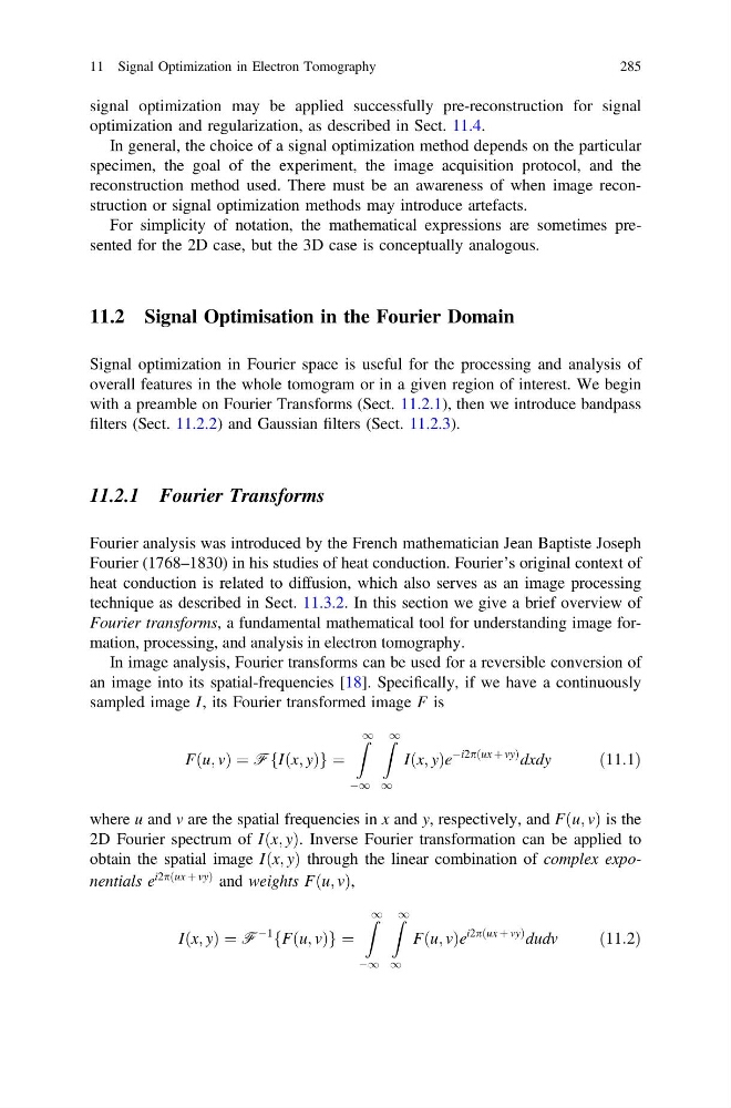
In image analysis, Fourier transforms can be used for a reversible conversion of
an image into its spatial-frequencies [18]. Specifically, if we have a continuously
sampled image I, its Fourier transformed image F is
Fðu; vÞ ¼ FfIðx; yÞg ¼
Z 1
À1
Z 1
1
Iðx; yÞe
Ài2pðux þ vyÞ dxdy
ð11:1Þ
where u and v are the spatial frequencies in x and y, respectively, and Fðu; vÞ is the
2D Fourier spectrum of Iðx; yÞ. Inverse Fourier transformation can be applied to
obtain the spatial image Iðx; yÞ through the linear combination of complex exponentials e
i2pðux þ vyÞ and weights Fðu; vÞ,
Iðx; yÞ ¼ F
À1
fFðu; vÞg ¼
Z 1
À1
Z 1
1
Fðu; vÞe
i2pðux þ vyÞ dudv
ð11:2Þ
11 Signal Optimization in Electron Tomography
285
optimization and regularization, as described in Sect. 11.4.
In general, the choice of a signal optimization method depends on the particular
specimen, the goal of the experiment, the image acquisition protocol, and the
reconstruction method used. There must be an awareness of when image reconstruction or signal optimization methods may introduce artefacts.
For simplicity of notation, the mathematical expressions are sometimes presented for the 2D case, but the 3D case is conceptually analogous.
11.2 Signal Optimisation in the Fourier Domain
Signal optimization in Fourier space is useful for the processing and analysis of
overall features in the whole tomogram or in a given region of interest. We begin
with a preamble on Fourier Transforms (Sect. 11.2.1), then we introduce bandpass
filters (Sect. 11.2.2) and Gaussian filters (Sect. 11.2.3).
11.2.1 Fourier Transforms
Fourier analysis was introduced by the French mathematician Jean Baptiste Joseph
Fourier (1768–1830) in his studies of heat conduction. Fourier’s original context of
heat conduction is related to diffusion, which also serves as an image processing
technique as described in Sect. 11.3.2. In this section we give a brief overview of
Fourier transforms, a fundamental mathematical tool for understanding image formation, processing, and analysis in electron tomography.
In image analysis, Fourier transforms can be used for a reversible conversion of
an image into its spatial-frequencies [18]. Specifically, if we have a continuously
sampled image I, its Fourier transformed image F is
Fðu; vÞ ¼ FfIðx; yÞg ¼
Z 1
À1
Z 1
1
Iðx; yÞe
Ài2pðux þ vyÞ dxdy
ð11:1Þ
where u and v are the spatial frequencies in x and y, respectively, and Fðu; vÞ is the
2D Fourier spectrum of Iðx; yÞ. Inverse Fourier transformation can be applied to
obtain the spatial image Iðx; yÞ through the linear combination of complex exponentials e
i2pðux þ vyÞ and weights Fðu; vÞ,
Iðx; yÞ ¼ F
À1
fFðu; vÞg ¼
Z 1
À1
Z 1
1
Fðu; vÞe
i2pðux þ vyÞ dudv
ð11:2Þ
11 Signal Optimization in Electron Tomography
285