

a picture from a finite subset of its projections taken at distinct angles.” Completeness
of information, however, depends on sampling statistics. For cryo- electron tomography, this is an apparent paradox. The requirement for as many projections as possible while covering the full angular range conflicts with the need to keep the electron
dose at subcritical levels to avoid erasing details. This means that a finite electron dose
is typically fractionated over the available tilt range. The “dose fractionation theorem”
[74, 75] states that the dose tolerated by the specimen can be divided by the number of
contributing projections to provide the maximum permissible dose for any one projection image of the tilt series. A negligible proportion of the dose is expended on
secondary tasks such as searching, tracking, and focussing, and ideally, tracking and
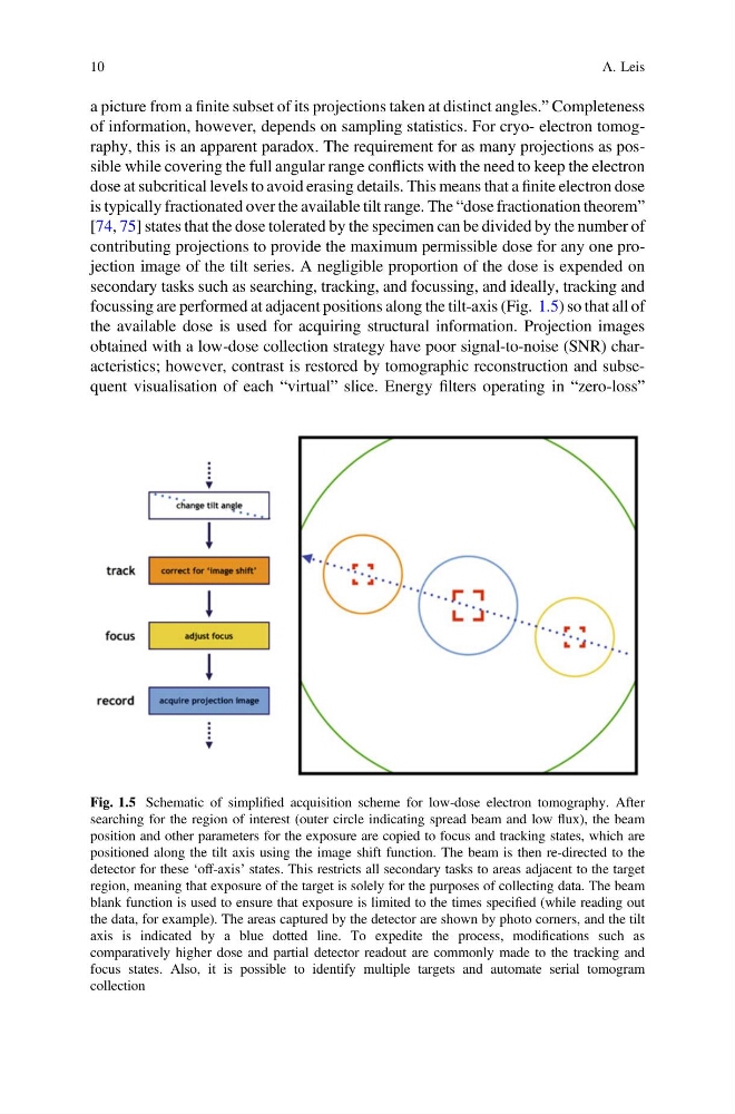
focussing are performed at adjacent positions along the tilt-axis (Fig. 1.5) so that all of
the available dose is used for acquiring structural information. Projection images
obtained with a low-dose collection strategy have poor signal-to-noise (SNR) characteristics; however, contrast is restored by tomographic reconstruction and subsequent visualisation of each “virtual” slice. Energy filters operating in “zero-loss”
Fig. 1.5 Schematic of simplified acquisition scheme for low-dose electron tomography. After
searching for the region of interest (outer circle indicating spread beam and low flux), the beam
position and other parameters for the exposure are copied to focus and tracking states, which are
positioned along the tilt axis using the image shift function. The beam is then re-directed to the
detector for these ‘off-axis’ states. This restricts all secondary tasks to areas adjacent to the target
region, meaning that exposure of the target is solely for the purposes of collecting data. The beam
blank function is used to ensure that exposure is limited to the times specified (while reading out
the data, for example). The areas captured by the detector are shown by photo corners, and the tilt
axis is indicated by a blue dotted line. To expedite the process, modifications such as
comparatively higher dose and partial detector readout are commonly made to the tracking and
focus states. Also, it is possible to identify multiple targets and automate serial tomogram
collection
10
A. Leis
of information, however, depends on sampling statistics. For cryo- electron tomography, this is an apparent paradox. The requirement for as many projections as possible while covering the full angular range conflicts with the need to keep the electron
dose at subcritical levels to avoid erasing details. This means that a finite electron dose
is typically fractionated over the available tilt range. The “dose fractionation theorem”
[74, 75] states that the dose tolerated by the specimen can be divided by the number of
contributing projections to provide the maximum permissible dose for any one projection image of the tilt series. A negligible proportion of the dose is expended on
secondary tasks such as searching, tracking, and focussing, and ideally, tracking and
focussing are performed at adjacent positions along the tilt-axis (Fig. 1.5) so that all of
the available dose is used for acquiring structural information. Projection images
obtained with a low-dose collection strategy have poor signal-to-noise (SNR) characteristics; however, contrast is restored by tomographic reconstruction and subsequent visualisation of each “virtual” slice. Energy filters operating in “zero-loss”
Fig. 1.5 Schematic of simplified acquisition scheme for low-dose electron tomography. After
searching for the region of interest (outer circle indicating spread beam and low flux), the beam
position and other parameters for the exposure are copied to focus and tracking states, which are
positioned along the tilt axis using the image shift function. The beam is then re-directed to the
detector for these ‘off-axis’ states. This restricts all secondary tasks to areas adjacent to the target
region, meaning that exposure of the target is solely for the purposes of collecting data. The beam
blank function is used to ensure that exposure is limited to the times specified (while reading out
the data, for example). The areas captured by the detector are shown by photo corners, and the tilt
axis is indicated by a blue dotted line. To expedite the process, modifications such as
comparatively higher dose and partial detector readout are commonly made to the tracking and
focus states. Also, it is possible to identify multiple targets and automate serial tomogram
collection
10
A. Leis