

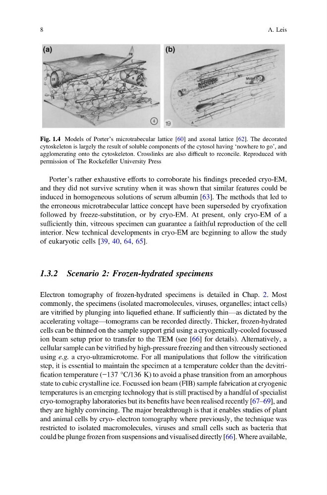
Porter’s rather exhaustive efforts to corroborate his findings preceded cryo-EM,
and they did not survive scrutiny when it was shown that similar features could be
induced in homogeneous solutions of serum albumin [63]. The methods that led to
the erroneous microtrabecular lattice concept have been superseded by cryofixation
followed by freeze-substitution, or by cryo-EM. At present, only cryo-EM of a
sufficiently thin, vitreous specimen can guarantee a faithful reproduction of the cell
interior. New technical developments in cryo-EM are beginning to allow the study
of eukaryotic cells [39, 40, 64, 65].
1.3.2 Scenario 2: Frozen-hydrated specimens
Electron tomography of frozen-hydrated specimens is detailed in Chap. 2. Most
commonly, the specimens (isolated macromolecules, viruses, organelles; intact cells)
are vitrified by plunging into liquefied ethane. If sufficiently thin—as dictated by the
accelerating voltage—tomograms can be recorded directly. Thicker, frozen-hydrated
cells can be thinned on the sample support grid using a cryogenically-cooled focussed
ion beam setup prior to transfer to the TEM (see [66] for details). Alternatively, a
cellular sample can be vitrified by high-pressure freezing and then vitreously sectioned
using e.g. a cryo-ultramicrotome. For all manipulations that follow the vitrification
step, it is essential to maintain the specimen at a temperature colder than the devitrification temperature (−137 °C/136 K) to avoid a phase transition from an amorphous
state to cubic crystalline ice. Focussed ion beam (FIB) sample fabrication at cryogenic
temperatures is an emerging technology that is still practised by a handful of specialist
cryo-tomography laboratories but its benefits have been realised recently [67–69], and
they are highly convincing. The major breakthrough is that it enables studies of plant
and animal cells by cryo- electron tomography where previously, the technique was
restricted to isolated macromolecules, viruses and small cells such as bacteria that
could be plunge frozen from suspensions and visualised directly [66]. Where available,
Fig. 1.4 Models of Porter’s microtrabecular lattice [60] and axonal lattice [62]. The decorated
cytoskeleton is largely the result of soluble components of the cytosol having ‘nowhere to go’, and
agglomerating onto the cytoskeleton. Crosslinks are also difficult to reconcile. Reproduced with
permission of The Rockefeller University Press
8
A. Leis
and they did not survive scrutiny when it was shown that similar features could be
induced in homogeneous solutions of serum albumin [63]. The methods that led to
the erroneous microtrabecular lattice concept have been superseded by cryofixation
followed by freeze-substitution, or by cryo-EM. At present, only cryo-EM of a
sufficiently thin, vitreous specimen can guarantee a faithful reproduction of the cell
interior. New technical developments in cryo-EM are beginning to allow the study
of eukaryotic cells [39, 40, 64, 65].
1.3.2 Scenario 2: Frozen-hydrated specimens
Electron tomography of frozen-hydrated specimens is detailed in Chap. 2. Most
commonly, the specimens (isolated macromolecules, viruses, organelles; intact cells)
are vitrified by plunging into liquefied ethane. If sufficiently thin—as dictated by the
accelerating voltage—tomograms can be recorded directly. Thicker, frozen-hydrated
cells can be thinned on the sample support grid using a cryogenically-cooled focussed
ion beam setup prior to transfer to the TEM (see [66] for details). Alternatively, a
cellular sample can be vitrified by high-pressure freezing and then vitreously sectioned
using e.g. a cryo-ultramicrotome. For all manipulations that follow the vitrification
step, it is essential to maintain the specimen at a temperature colder than the devitrification temperature (−137 °C/136 K) to avoid a phase transition from an amorphous
state to cubic crystalline ice. Focussed ion beam (FIB) sample fabrication at cryogenic
temperatures is an emerging technology that is still practised by a handful of specialist
cryo-tomography laboratories but its benefits have been realised recently [67–69], and
they are highly convincing. The major breakthrough is that it enables studies of plant
and animal cells by cryo- electron tomography where previously, the technique was
restricted to isolated macromolecules, viruses and small cells such as bacteria that
could be plunge frozen from suspensions and visualised directly [66]. Where available,
Fig. 1.4 Models of Porter’s microtrabecular lattice [60] and axonal lattice [62]. The decorated
cytoskeleton is largely the result of soluble components of the cytosol having ‘nowhere to go’, and
agglomerating onto the cytoskeleton. Crosslinks are also difficult to reconcile. Reproduced with
permission of The Rockefeller University Press
8
A. Leis