

reconstruction approaches. de Rosier and Klug also mentioned the idea of a tilt
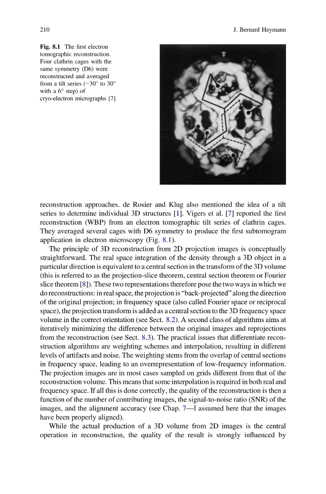
series to determine individual 3D structures [1]. Vigers et al. [7] reported the first
reconstruction (WBP) from an electron tomographic tilt series of clathrin cages.
They averaged several cages with D6 symmetry to produce the first subtomogram
application in electron microscopy (Fig. 8.1).
The principle of 3D reconstruction from 2D projection images is conceptually
straightforward. The real space integration of the density through a 3D object in a
particular direction is equivalent to a central section in the transform of the 3D volume
(this is referred to as the projection-slice theorem, central section theorem or Fourier
slice theorem [8]). These two representations therefore pose the two ways in which we
do reconstructions: in real space, the projection is “back-projected” along the direction
of the original projection; in frequency space (also called Fourier space or reciprocal
space), the projection transform is added as a central section to the 3D frequency space
volume in the correct orientation (see Sect. 8.2). A second class of algorithms aims at
iteratively minimizing the difference between the original images and reprojections
from the reconstruction (see Sect. 8.3). The practical issues that differentiate reconstruction algorithms are weighting schemes and interpolation, resulting in different
levels of artifacts and noise. The weighting stems from the overlap of central sections
in frequency space, leading to an overrepresentation of low-frequency information.
The projection images are in most cases sampled on grids different from that of the
reconstruction volume. This means that some interpolation is required in both real and
frequency space. If all this is done correctly, the quality of the reconstruction is then a
function of the number of contributing images, the signal-to-noise ratio (SNR) of the
images, and the alignment accuracy (see Chap. 7—I assumed here that the images
have been properly aligned).
While the actual production of a 3D volume from 2D images is the central
operation in reconstruction, the quality of the result is strongly influenced by
Fig. 8.1 The first electron
tomographic reconstruction.
Four clathrin cages with the
same symmetry (D6) were
reconstructed and averaged
from a tilt series (−30° to 30°
with a 6° step) of
cryo-electron micrographs [7]
210
J. Bernard Heymann
series to determine individual 3D structures [1]. Vigers et al. [7] reported the first
reconstruction (WBP) from an electron tomographic tilt series of clathrin cages.
They averaged several cages with D6 symmetry to produce the first subtomogram
application in electron microscopy (Fig. 8.1).
The principle of 3D reconstruction from 2D projection images is conceptually
straightforward. The real space integration of the density through a 3D object in a
particular direction is equivalent to a central section in the transform of the 3D volume
(this is referred to as the projection-slice theorem, central section theorem or Fourier
slice theorem [8]). These two representations therefore pose the two ways in which we
do reconstructions: in real space, the projection is “back-projected” along the direction
of the original projection; in frequency space (also called Fourier space or reciprocal
space), the projection transform is added as a central section to the 3D frequency space
volume in the correct orientation (see Sect. 8.2). A second class of algorithms aims at
iteratively minimizing the difference between the original images and reprojections
from the reconstruction (see Sect. 8.3). The practical issues that differentiate reconstruction algorithms are weighting schemes and interpolation, resulting in different
levels of artifacts and noise. The weighting stems from the overlap of central sections
in frequency space, leading to an overrepresentation of low-frequency information.
The projection images are in most cases sampled on grids different from that of the
reconstruction volume. This means that some interpolation is required in both real and
frequency space. If all this is done correctly, the quality of the reconstruction is then a
function of the number of contributing images, the signal-to-noise ratio (SNR) of the
images, and the alignment accuracy (see Chap. 7—I assumed here that the images
have been properly aligned).
While the actual production of a 3D volume from 2D images is the central
operation in reconstruction, the quality of the result is strongly influenced by
Fig. 8.1 The first electron
tomographic reconstruction.
Four clathrin cages with the
same symmetry (D6) were
reconstructed and averaged
from a tilt series (−30° to 30°
with a 6° step) of
cryo-electron micrographs [7]
210
J. Bernard Heymann