

and the similarities of many complexes at the best, attainable resolution, it can be more
difficult to assign structure or determine interactions without explicit, a priori information. Therefore, this group of techniques should be considered together as a
complementary and powerful means of determining structure-function relationships
in cells [12]. Correlative light and electron microscopy (CLEM, [13–17]) seeks to
apply such technologies in combination for the elucidation of structure-function
relationships.
From an instrumentation perspective, electron tomography belongs to the group
of electron microscope -based technologies capable of generating 3D structures,
collectively referred to as ‘3D electron microscopy’ (3DEM, [18]). 3DEM comprises
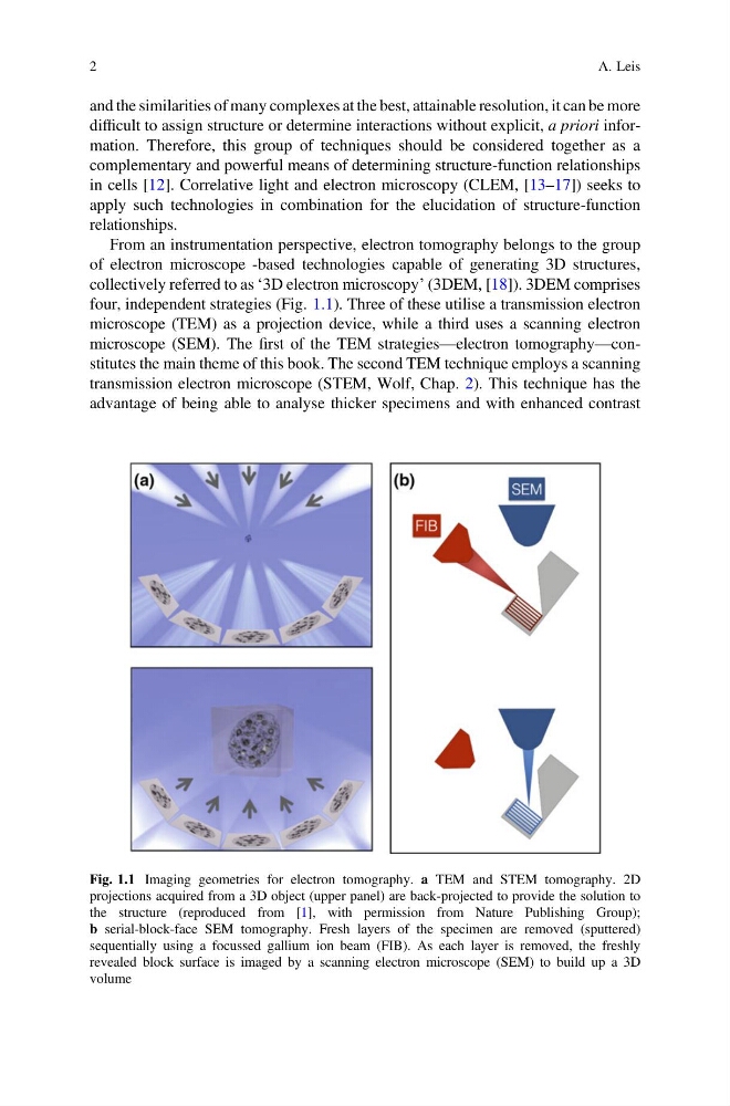
four, independent strategies (Fig. 1.1). Three of these utilise a transmission electron
microscope (TEM) as a projection device, while a third uses a scanning electron
microscope (SEM). The first of the TEM strategies—electron tomography—constitutes the main theme of this book. The second TEM technique employs a scanning
transmission electron microscope (STEM, Wolf, Chap. 2). This technique has the
advantage of being able to analyse thicker specimens and with enhanced contrast
Fig. 1.1 Imaging geometries for electron tomography. a TEM and STEM tomography. 2D
projections acquired from a 3D object (upper panel) are back-projected to provide the solution to
the structure (reproduced from [1], with permission from Nature Publishing Group);
b serial-block-face SEM tomography. Fresh layers of the specimen are removed (sputtered)
sequentially using a focussed gallium ion beam (FIB). As each layer is removed, the freshly
revealed block surface is imaged by a scanning electron microscope (SEM) to build up a 3D
volume
2
A. Leis
difficult to assign structure or determine interactions without explicit, a priori information. Therefore, this group of techniques should be considered together as a
complementary and powerful means of determining structure-function relationships
in cells [12]. Correlative light and electron microscopy (CLEM, [13–17]) seeks to
apply such technologies in combination for the elucidation of structure-function
relationships.
From an instrumentation perspective, electron tomography belongs to the group
of electron microscope -based technologies capable of generating 3D structures,
collectively referred to as ‘3D electron microscopy’ (3DEM, [18]). 3DEM comprises
four, independent strategies (Fig. 1.1). Three of these utilise a transmission electron
microscope (TEM) as a projection device, while a third uses a scanning electron
microscope (SEM). The first of the TEM strategies—electron tomography—constitutes the main theme of this book. The second TEM technique employs a scanning
transmission electron microscope (STEM, Wolf, Chap. 2). This technique has the
advantage of being able to analyse thicker specimens and with enhanced contrast
Fig. 1.1 Imaging geometries for electron tomography. a TEM and STEM tomography. 2D
projections acquired from a 3D object (upper panel) are back-projected to provide the solution to
the structure (reproduced from [1], with permission from Nature Publishing Group);
b serial-block-face SEM tomography. Fresh layers of the specimen are removed (sputtered)
sequentially using a focussed gallium ion beam (FIB). As each layer is removed, the freshly
revealed block surface is imaged by a scanning electron microscope (SEM) to build up a 3D
volume
2
A. Leis