

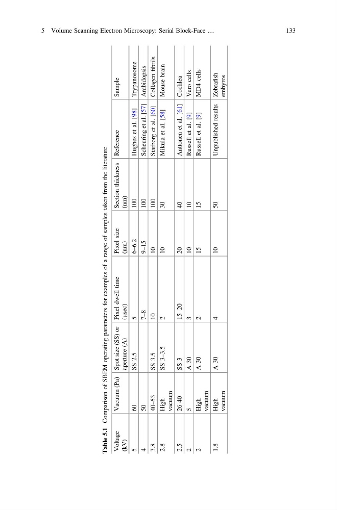
Table 5.1
Comparison of SBEM operating parameters for examples of a range of samples taken from the literature
Voltage
(kV)
Vacuum (Pa) Spot size (SS) or
aperture (A)
Pixel dwell time
(µsec)
Pixel size
(nm)
Section thickness
(nm)
Reference
Sample
5
60
SS 2.5
5
6–6.2
100
Hughes et al. [98]
Trypanosome
4
5 0
7
–8
9
–15
100
Scheuring et al. [57] Arabidopsis
3.8
40–53
SS 3.5
10
10
100
Starborg et al. [60] Collagen
fibrils
2.8
High
vacuum
SS 3–3.5
2
10
30
Mikula et al. [58]
Mouse brain
2.5
26-40
SS 3
15–20
20
40
Anttonen et al. [61] Cochlea
2
5
A 30
3
10
10
Russell et al. [9]
Vero cells
2
High
vacuum
A 30
2
15
15
Russell et al. [9]
MD4 cells
1.8
High
vacuum
A 30
4
10
50
Unpublished results Zebrafish
embyros
5 Volume Scanning Electron Microscopy: Serial Block-Face …
133
Comparison of SBEM operating parameters for examples of a range of samples taken from the literature
Voltage
(kV)
Vacuum (Pa) Spot size (SS) or
aperture (A)
Pixel dwell time
(µsec)
Pixel size
(nm)
Section thickness
(nm)
Reference
Sample
5
60
SS 2.5
5
6–6.2
100
Hughes et al. [98]
Trypanosome
4
5 0
7
–8
9
–15
100
Scheuring et al. [57] Arabidopsis
3.8
40–53
SS 3.5
10
10
100
Starborg et al. [60] Collagen
fibrils
2.8
High
vacuum
SS 3–3.5
2
10
30
Mikula et al. [58]
Mouse brain
2.5
26-40
SS 3
15–20
20
40
Anttonen et al. [61] Cochlea
2
5
A 30
3
10
10
Russell et al. [9]
Vero cells
2
High
vacuum
A 30
2
15
15
Russell et al. [9]
MD4 cells
1.8
High
vacuum
A 30
4
10
50
Unpublished results Zebrafish
embyros
5 Volume Scanning Electron Microscopy: Serial Block-Face …
133