

the vacuum). At low magnification the sample is searched to find the ROI. It is
advisable to work at low voltage, though sometimes a higher voltage can be useful
for revealing the sample anatomy below the block-face. When working with cell
cultures that were embedded on the culture plate, a voltage of 5 kV will make it
possible to view the cells below the surface of the resin, although this will cause
some charging. Once more detailed viewing is needed and the magnification is
increased, the microscope and SBEM will need to be setup so as not to damage the
sample.
The structure of interest may not be in view immediately and it may take some
sectioning and continuous checking until the region is reached. In order to find the
ROI and capture a data set of the entire structure it is necessary to use neighbouring
markers to locate it before sectioning commences.
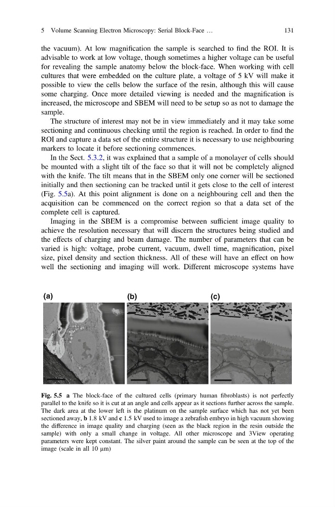
In the Sect. 5.3.2, it was explained that a sample of a monolayer of cells should
be mounted with a slight tilt of the face so that it will not be completely aligned
with the knife. The tilt means that in the SBEM only one corner will be sectioned
initially and then sectioning can be tracked until it gets close to the cell of interest
(Fig. 5.5a). At this point alignment is done on a neighbouring cell and then the
acquisition can be commenced on the correct region so that a data set of the
complete cell is captured.
Imaging in the SBEM is a compromise between sufficient image quality to
achieve the resolution necessary that will discern the structures being studied and
the effects of charging and beam damage. The number of parameters that can be
varied is high: voltage, probe current, vacuum, dwell time, magnification, pixel
size, pixel density and section thickness. All of these will have an effect on how
well the sectioning and imaging will work. Different microscope systems have
(a)
(b)
(c)
Fig. 5.5 a The block-face of the cultured cells (primary human fibroblasts) is not perfectly
parallel to the knife so it is cut at an angle and cells appear as it sections further across the sample.
The dark area at the lower left is the platinum on the sample surface which has not yet been
sectioned away, b 1.8 kV and c 1.5 kV used to image a zebrafish embryo in high vacuum showing
the difference in image quality and charging (seen as the black region in the resin outside the
sample) with only a small change in voltage. All other microscope and 3View operating
parameters were kept constant. The silver paint around the sample can be seen at the top of the
image (scale in all 10 µm)
5 Volume Scanning Electron Microscopy: Serial Block-Face …
131
advisable to work at low voltage, though sometimes a higher voltage can be useful
for revealing the sample anatomy below the block-face. When working with cell
cultures that were embedded on the culture plate, a voltage of 5 kV will make it
possible to view the cells below the surface of the resin, although this will cause
some charging. Once more detailed viewing is needed and the magnification is
increased, the microscope and SBEM will need to be setup so as not to damage the
sample.
The structure of interest may not be in view immediately and it may take some
sectioning and continuous checking until the region is reached. In order to find the
ROI and capture a data set of the entire structure it is necessary to use neighbouring
markers to locate it before sectioning commences.
In the Sect. 5.3.2, it was explained that a sample of a monolayer of cells should
be mounted with a slight tilt of the face so that it will not be completely aligned
with the knife. The tilt means that in the SBEM only one corner will be sectioned
initially and then sectioning can be tracked until it gets close to the cell of interest
(Fig. 5.5a). At this point alignment is done on a neighbouring cell and then the
acquisition can be commenced on the correct region so that a data set of the
complete cell is captured.
Imaging in the SBEM is a compromise between sufficient image quality to
achieve the resolution necessary that will discern the structures being studied and
the effects of charging and beam damage. The number of parameters that can be
varied is high: voltage, probe current, vacuum, dwell time, magnification, pixel
size, pixel density and section thickness. All of these will have an effect on how
well the sectioning and imaging will work. Different microscope systems have
(a)
(b)
(c)
Fig. 5.5 a The block-face of the cultured cells (primary human fibroblasts) is not perfectly
parallel to the knife so it is cut at an angle and cells appear as it sections further across the sample.
The dark area at the lower left is the platinum on the sample surface which has not yet been
sectioned away, b 1.8 kV and c 1.5 kV used to image a zebrafish embryo in high vacuum showing
the difference in image quality and charging (seen as the black region in the resin outside the
sample) with only a small change in voltage. All other microscope and 3View operating
parameters were kept constant. The silver paint around the sample can be seen at the top of the
image (scale in all 10 µm)
5 Volume Scanning Electron Microscopy: Serial Block-Face …
131