

high signal-to-noise ratio (SNR) by producing high numbers of backscattered
electrons from the heavy metal stained structures. Lateral resolution is also
important and this is a factor of voltage and probe size. However, when deciding on
the imaging conditions, it is a balance between image quality, acquisition speed,
sample damage and the desired results.
During a 3D acquisition, in both SBEM and FIB-SEM, the images are acquired
using a backscattered electron detector that sits above the sample. Using this form
of imaging, contrast is given by light elements producing dark coloured pixels and
heavy elements producing bright pixels. As images of this type are foreign to most
biologists they are inverted to produce an image that looks similar to a TEM image.
In order to reduce charging in the sample, the SEM is operated at low voltages
(usually lower than 3 kV) for imaging during 3D acquisitions. Lower kVs also
ensure the signal is coming from the surface or only a short distance below. The
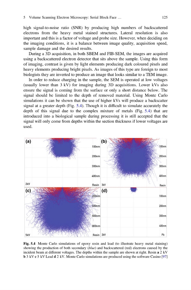
signal should be limited to the depth of removed material. Using Monte Carlo
simulations it can be shown that the use of higher kVs will produce a backscatter
signal at a greater depth (Fig. 5.4). Though it is difficult to simulate accurately the
depth of this signal due to the complex mixture of metals (Fig. 5.4) that are
introduced into a biological sample during processing it is still accepted that the
signal will only come from depths within the section thickness if lower voltages are
used.
Fig. 5.4 Monte Carlo simulations of epoxy resin and lead (to illustrate heavy metal staining)
showing the production of both secondary (blue) and backscattered (red) electrons caused by the
incident beam at different voltages. The depths within the sample are shown at right. Resin a 2 kV
b 3 kV c 5 kV Lead d 2 kV. Monte Carlo simulations are produced using the software Casino [97]
5 Volume Scanning Electron Microscopy: Serial Block-Face …
125
electrons from the heavy metal stained structures. Lateral resolution is also
important and this is a factor of voltage and probe size. However, when deciding on
the imaging conditions, it is a balance between image quality, acquisition speed,
sample damage and the desired results.
During a 3D acquisition, in both SBEM and FIB-SEM, the images are acquired
using a backscattered electron detector that sits above the sample. Using this form
of imaging, contrast is given by light elements producing dark coloured pixels and
heavy elements producing bright pixels. As images of this type are foreign to most
biologists they are inverted to produce an image that looks similar to a TEM image.
In order to reduce charging in the sample, the SEM is operated at low voltages
(usually lower than 3 kV) for imaging during 3D acquisitions. Lower kVs also
ensure the signal is coming from the surface or only a short distance below. The
signal should be limited to the depth of removed material. Using Monte Carlo
simulations it can be shown that the use of higher kVs will produce a backscatter
signal at a greater depth (Fig. 5.4). Though it is difficult to simulate accurately the
depth of this signal due to the complex mixture of metals (Fig. 5.4) that are
introduced into a biological sample during processing it is still accepted that the
signal will only come from depths within the section thickness if lower voltages are
used.
Fig. 5.4 Monte Carlo simulations of epoxy resin and lead (to illustrate heavy metal staining)
showing the production of both secondary (blue) and backscattered (red) electrons caused by the
incident beam at different voltages. The depths within the sample are shown at right. Resin a 2 kV
b 3 kV c 5 kV Lead d 2 kV. Monte Carlo simulations are produced using the software Casino [97]
5 Volume Scanning Electron Microscopy: Serial Block-Face …
125