

Particularly for SBEM, sample processing is an integral part of the technique
largely to avoid charging problems. The development of a processing protocol that
gave more consistent results [41, 42] helped to project SBEM into becoming a
routine technique. This protocol has been designed to deposit large amounts of
(a)
(b)
(d)
(e)
(c)
(f)
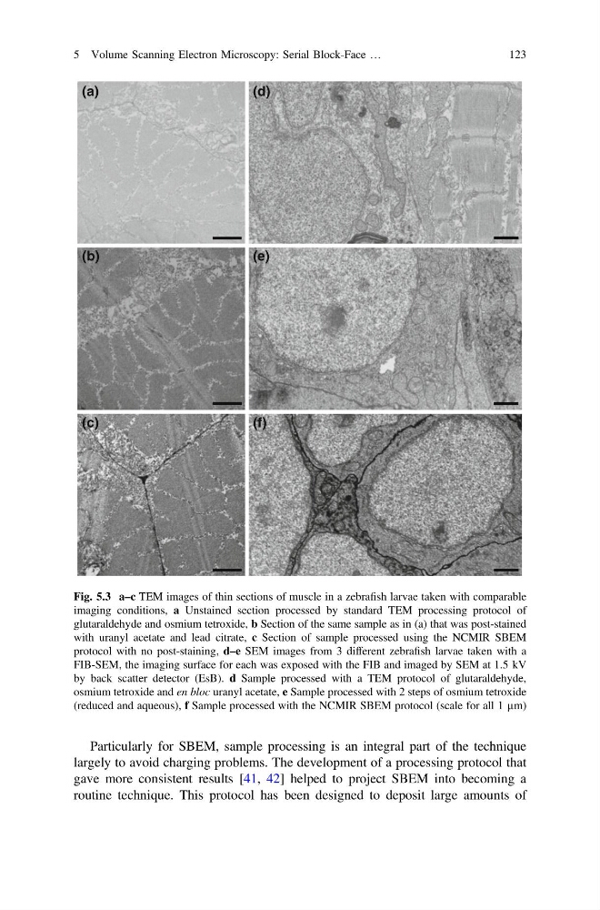
Fig. 5.3 a–c TEM images of thin sections of muscle in a zebrafish larvae taken with comparable
imaging conditions, a Unstained section processed by standard TEM processing protocol of
glutaraldehyde and osmium tetroxide, b Section of the same sample as in (a) that was post-stained
with uranyl acetate and lead citrate, c Section of sample processed using the NCMIR SBEM
protocol with no post-staining, d–e SEM images from 3 different zebrafish larvae taken with a
FIB-SEM, the imaging surface for each was exposed with the FIB and imaged by SEM at 1.5 kV
by back scatter detector (EsB). d Sample processed with a TEM protocol of glutaraldehyde,
osmium tetroxide and en bloc uranyl acetate, e Sample processed with 2 steps of osmium tetroxide
(reduced and aqueous), f Sample processed with the NCMIR SBEM protocol (scale for all 1 µm)
5 Volume Scanning Electron Microscopy: Serial Block-Face …
123
largely to avoid charging problems. The development of a processing protocol that
gave more consistent results [41, 42] helped to project SBEM into becoming a
routine technique. This protocol has been designed to deposit large amounts of
(a)
(b)
(d)
(e)
(c)
(f)
Fig. 5.3 a–c TEM images of thin sections of muscle in a zebrafish larvae taken with comparable
imaging conditions, a Unstained section processed by standard TEM processing protocol of
glutaraldehyde and osmium tetroxide, b Section of the same sample as in (a) that was post-stained
with uranyl acetate and lead citrate, c Section of sample processed using the NCMIR SBEM
protocol with no post-staining, d–e SEM images from 3 different zebrafish larvae taken with a
FIB-SEM, the imaging surface for each was exposed with the FIB and imaged by SEM at 1.5 kV
by back scatter detector (EsB). d Sample processed with a TEM protocol of glutaraldehyde,
osmium tetroxide and en bloc uranyl acetate, e Sample processed with 2 steps of osmium tetroxide
(reduced and aqueous), f Sample processed with the NCMIR SBEM protocol (scale for all 1 µm)
5 Volume Scanning Electron Microscopy: Serial Block-Face …
123